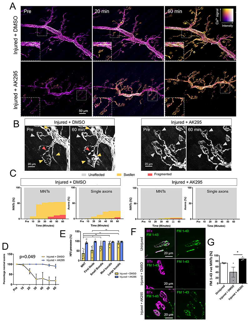
Fig. 5. Calpain inhibition prevents adverse morphological changes in the axon without affecting calcium influx.
A: Ratiometric calcium images from maximum intensity projections showing axonal calcium at different timepoints. Colour key demonstrates relative calcium levels, with “hotter” colours indicating higher calcium and brightness indicating fluorescent intensity. Insets show changes at individual MNTs. B: Maximum intensity projections of single channel showing morphological changes at MNT in explants which were injured using Ab+NHS with the addition of either AK295 or DMSO vehicle control. Grey arrows = unaffected axons, yellow arrows = swollen axons and red = fragmented axons. C: Quantification of percentages of unaffected, swollen and fragmented axons at the MNT and single axons in DMSO or AK295 treated injured explants. D: Morphological changes seen in large bundles (p = 0.049 two-way repeated measures ANOVA with Sidak’s multiple comparisons test). n = 4 Injured + DMSO, n = 3 Injured + AK295 E: Quantification of NFil (top) and AnkG (bottom) presence. # = p < 0.05, ## = vs Ab only (p < 0.01), two-way repeated measures ANOVA with Sidak’s multiple comparison’s test. * = p < 0.05, ** = p < 0.01, *** = p < 0.001, two-way repeated measure ANOVA with Tukey’s multiple comparisons test. n = 4 Injured+DMSO, n = 3 Injured+AK295 F: Illustrative images showing uptake of FM 1–43 at the MNTs of uninjured, injured + DMSO or injured + AK295 explants. Staining in both injured groups was more punctate than uninjured explants. G: Quantification of FM 1–43 presence at MNTs (p < 0.05, one-way ANOVA with Tukey’s multiple comparisons test). n = 3 for both groups. (For interpretation of the references to colour in this figure legend, the reader is referred to the web version of this article.)

