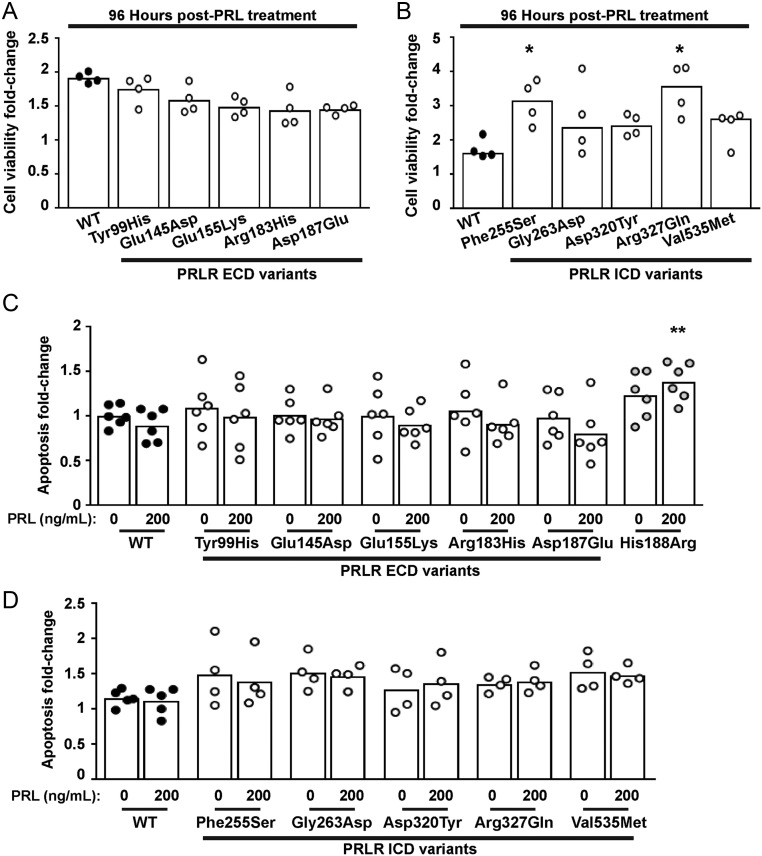
Figure 8.
Effect of the PRLR rare variants on cell viability and apoptosis. (A-B) Effect of PRL (200 ng/mL) on viability in cells expressing WT, or (A) the extracellular domain (ECD) or (B) the intracellular domain (ICD) variant PRLRs. Cell viability was increased in cells expressing the Phe255Ser and Arg327Gln variant PRLRs at 96 h post-treatment with PRL, when compared to WT cells. (C-D) Effect of PRL (200 ng/mL) on apoptosis in cells expressing WT, mutant His188Arg, or (C) the ECD or (D) the ICD variant PRLRs. Each point shows one biological replicate (derived from the mean of four technical replicates) performed on independent occasions. Statistical analyses performed by one-way ANOVA with Dunnett’s multiple comparisons tests for panels A, C, and D. Statistical analysis performed by Kruskal–Wallis test with Dunn’s multiple comparisons tests for panel B. Comparisons show WT vs variant (asterisks). ****P < 0.0001, ***P < 0.001, **P < 0.01, *P < 0.05.

 This work is licensed under a