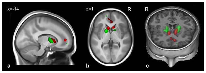
Figure 1.
Regions with smaller gray matter volume (red) and white matter volume (green) in 119 adolescents with subthreshold depression versus 461 controls. Note: voxel level was set at p < .05 (familywise error–corrected for the whole brain). Results are superimposed on a T1-weighted magnetic resonance imaging scan of an adolescent brain from the Imagen database. (a) Sagittal slice; (b) transversal slice; (c) 3-dimensional representation (coronal and transversal slices). R = right.

