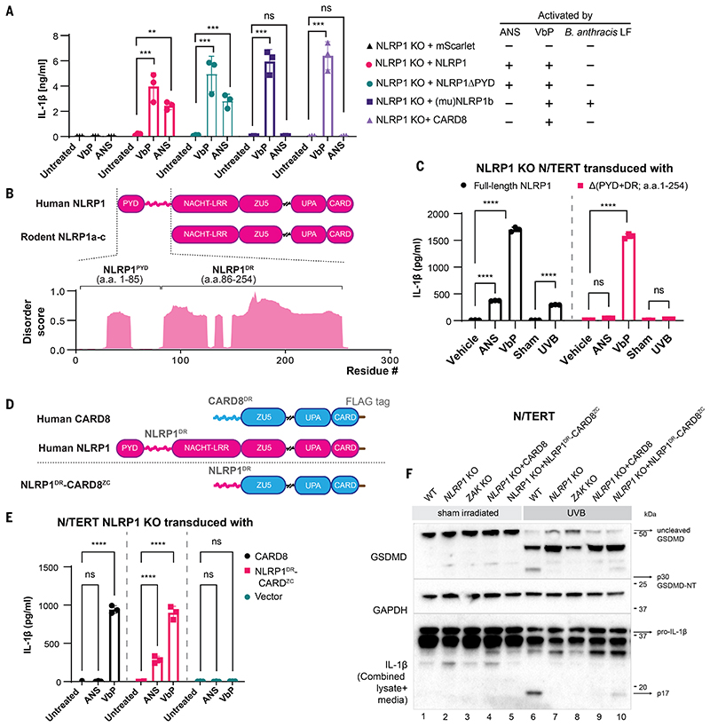
Figure 3. A disordered linker region of human NLRP1 selectively mediates ZAKα-dependent activation.
(A) IL-1β ELISA from NLRP1 KO N/TERT cells reconstituted with the indicated NLRP1 variants and CARD8 treated with ANS (1 μM) and VbP (3 μM). Note that this experiment was performed independently using higher cell numbers. (B) Comparison between the domain structures of human NLRP1 and rodent NLRP1a-c. The predicted disorder score was calculated for a.a. 1-300 of human NLRP1. (C) IL-1β ELISA from NLRP1-KO N/TERT cells reconstituted with GFP-full-length NLRP1 or NLRP1 lacking PYD+DR (a.a.1-254). Cells were treated with the indicated drugs, or sham- or UVB-irradiated and harvested 24 hours post-treatment. (D) Comparison of the domain arrangements of human NLRP1 and CARD8 and the engineered hybrid sensor referred to as NLRP1DR-CARD8ZC. (E) IL-1β ELISA from NLRP1-KO N/TERT cells transduced with CARD8 or NLRP1DR-CARD8ZC and treated with 1 μM ANS or 3 μM VbP for 24 hours. (F) GSDMD and IL-1β immunoblot from the cells in E, along with WT or ZAK KO N/TERT cells irradiated with UVB. The GSDMD antibody recognizes both full-length and cleaved forms, including p43 and p30. The IL-1β immunoblot was performed with samples that combined lysate and 10 times concentrated media. The error bars represent standard errors of the mean (S.E.M.) from three biological replicates, where one replicate refers to an independent seeding and treatment of the cells. The significance values in (A), (C) and (E) were calculated based on two-way ANOVA followed by Sidak’s test for multiple pairwise comparisons. Significance values were indicated as: ns, non-significant, **P<0.01, *** P<0.001, ****P<0.0001.

