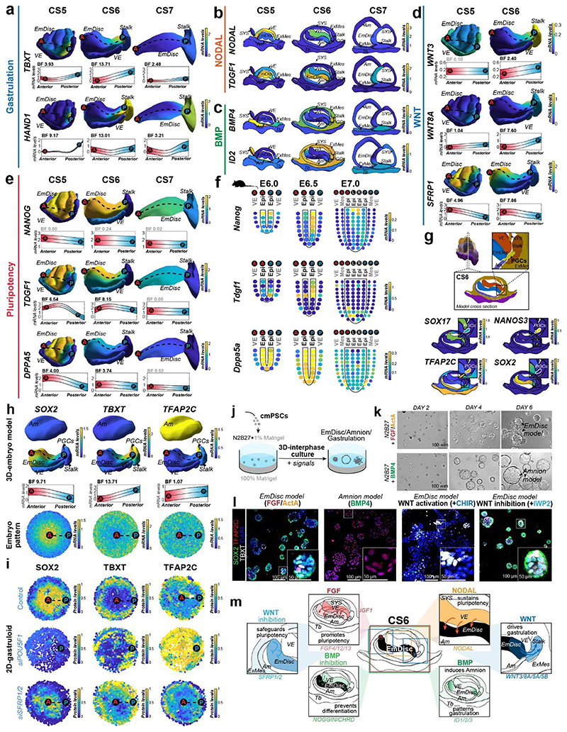
Figure 3. 3D-transcriptomes and stem cell-based embryo models delineate body axis formation.
a, GPR-based 3D-transcriptomes in EmDisc/VE showing gastrulation marker expression in the posterior EmDisc. Upper panels: Relative mRNA levels in EmDisc, VE, and stalk. Lower panels: mRNA expression change along anterior-posterior axis (dashed line; anterior (red, A) to posterior (blue, P)) in EmDisc, quantified by Bayesian factor (BF). b-c, Relative mRNA levels of (b) NODAL and (c) BMP in virtual embryo cross sections. d, WNT signalling pathway components shown in EmDisc/VE model. e, GPR-models for EmDisc/VE displaying regionalised pluripotency factor transcription in the anterior EmDisc. f, Spatial expression of pluripotency factors in gastrulating mouse embryos at E6.0, E6.5, and E7.0 according to Geo-seq30. g, Virtual cross-sections of the PGCs at CS6. GPR, Gaussian process regression; CS, Carnegie stage; EmDisc, Embryonic disc; VE, Visceral endoderm; ExMes, Extraembryonic Mesoderm. Am, Amnion; SYS, Secondary Yolk Sac; Tb, Trophoblast; PGC, Primordial Germ Cells. h, GPR-models for CS6 embryo of anterior marker (SOX2), posterior marker (TBXT) and amnion/PGC maker (TFAP2C). Upper panels: Relative mRNA levels for gene expression in EmDisc, VE, PGCs, Stalk, and Amnion. Am is displayed separately for visualisation. Middle panels: mRNA expression change along anterior-posterior axis (dashed line, anterior (red, A) to posterior (blue, P)) in EmDisc, quantified by Bayesian factor (BF). Lower panel: Virtual embryo pattern of EmDisc expression patterns generated by axis expression in middle panel. i, Expression patterns of in vitro 2D gastruloids segmented nuclei stained for anterior marker (SOX2), posterior marker (TBXT) and amnion/PGC maker (TFAP2C) after differentiation in BMP4 (50 ng/mL), FGF (10ng/mL), and Activin A (20ng/mL) under control conditions (top panel) and following siRNA transfection (bottom panels). Each intensity profile is normalized log expression levels are standardized so that they vary within [0,1]. j, Schematic representation of 3D-interphase culture system. cmPSCs are seeded on a bed of 100% Matrigel and overlaid with 1% Matrigel supplemented N2B27-based culture medium with and without signalling molecules. k, Brightfield images of EmDisc-like structures (N2B27 + 100 ng/mL FGF + 20 ng/mL Activin A) or Amnion-like structures (N2B27 + 50 ng/mL BMP4). l, Immunofluorescence images of structures generated after 4 days in EmDisc- or Amnion-promoting conditions or in EmDisc conditions with WNT modulation through 3 μM CHIR99021 (WNT activator) or 3 μM IWP-2 (WNT production inhibitor). m, Schematic summary diagram of BMP, WNT, FGF and NODAL signalling pathway activities in the marmoset embryo at CS6. PSCs, Pluripotent Stem Cells.

