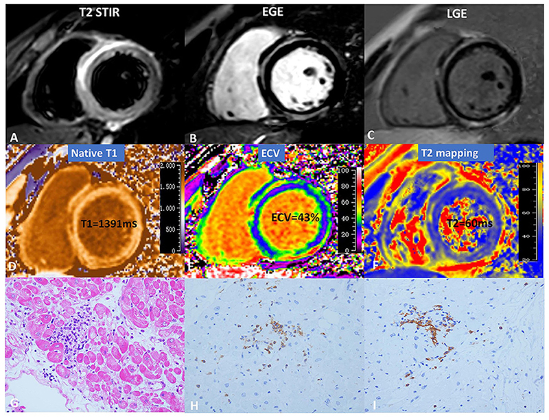
Figure 5. Cardiovascular magnetic resonance (CMR) and pathology results for a representative case of myocarditis.
(A) T2-STIR, (B) EGE, (C) LGE, (D) native T1, (E) ECV, and (F) T2-mapping. (G) indicates focal myocyte damage with lymphocytic infiltration. Immunohistochemistry revealed (H) LCA + (x40) and (I) CD20 + (x40). As originally published by Open Access Frontiers in Li et al. Frontiers in Cardiovascular Medicine. 2021;8:739892. T2 STIR = T2 short-tau inversion recovery, EGE = early gadolinium enhancement, LGE = late gadolinium enhancement, ECV = extracellular volume, LCA = leukocyte common antigen, CD = cluster of differentiation 70

