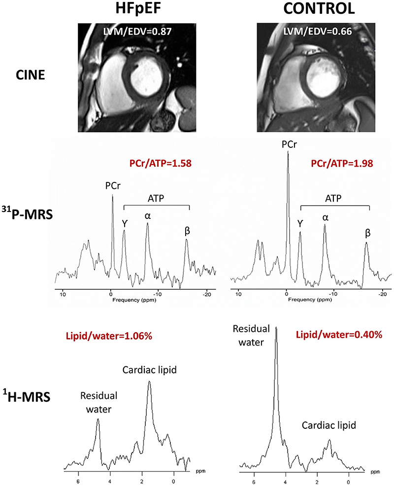
Figure 7.
Representative results of LVM/EDV, PCr/ATP and Lipid/water for heart failure with preserved ejection fraction (HFpEF) (left) and control (right). Cine imaging (top panel), 31P-CMRS (middle panel) and 1H-CMRS (bottom panel). 1H-CMRS spectra are scaled based on unsuppressed water (not shown) and noise level. LVM = left ventricular mass; EDV = end-diastolic volume; PCr = phosphocreatine; ATP = adenosine triphosphate; CMRS = cardiovascular magnetic resonance spectroscopy. As originally published by Springer Nature in Mahmod et al. Journal of Cardiovascular Magnetic Resonance. 2018;20(1):88 85

