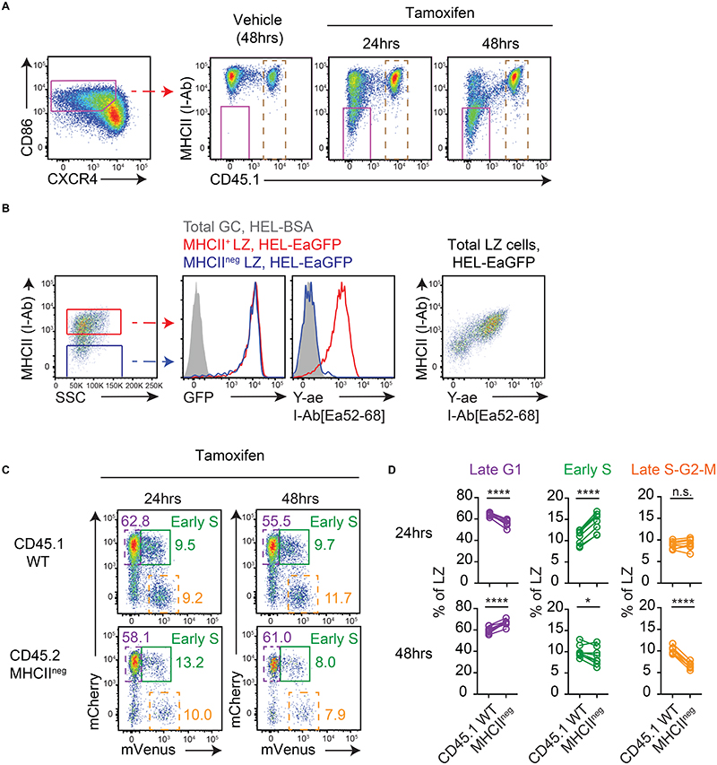
Figure 3. Cyclic re-entry initiation does not require MHCII presentation.
(A) Analysis of WT:R26-CreERT2 MHCIIfl/fl Fucci2 mixed BM chimeras on day 8 post-SRBC immunisation, at indicated times after tamoxifen treatment. Representative plots of MHCII deletion by LZ splenic GC B cells (IgDlow CD95+ GL7+). Dashed gates identify WT cells used in (C-D). (B) Antigen acquisition (GFP) and pMHCII presentation (Y-ae mAb staining) by day 8 SWHEL Cd79a-CreERT2 MHCIIfl/fl LZ GC B cells (40hrs post-tamoxifen) following 2 hr ex vivo incubations with HEL-EaGFP or HEL-BSA (control).(C) LZ populations gated in A. Frequencies of late G1 (dashed purple), early S (green) and late S-G2-M (dashed orange) cells determined for WT and MHCII-deleted populations, (D) and summarised. Lines join populations from individual mice. Results show representative FACs plots (A, B, C) or pooled results (D) from 2 experiments each containing 4-5 mice per condition. Analysis, paired two-tailed Student’s t test. *p < 0.05, ****p < 0.0001.

