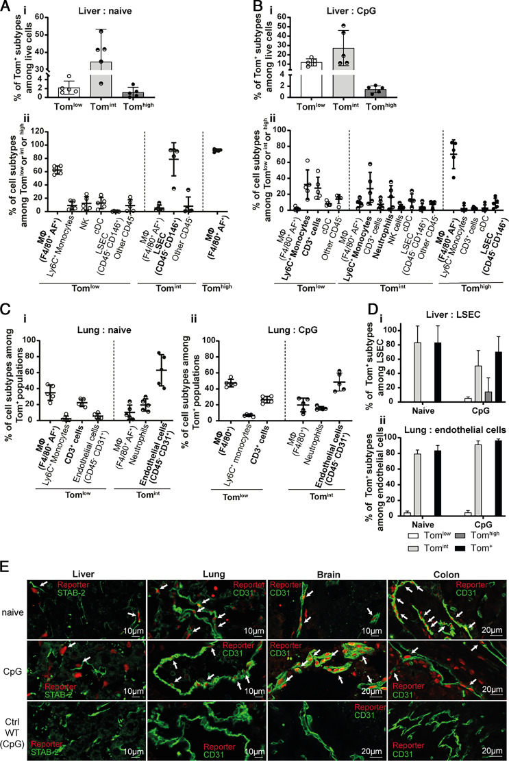
FIGURE 2.
Endothelial cells are a source of IL-18BP in various organs. (A, B) Flow cytometric analysis of Tom+ subpopulations in liver of (A) naive (n = 5) and (B) CpG-injected (n = 5) KI mice revealing (A-Bi) their percentage among live cells and (A-Bii) reporter-positive cell types. (C) Identification of Tomlow and Tomint cell types in the lung of (Ci) naive (n = 5) and (Cii) CpG-injected (n = 5) KI mice. (D) Percentage of each reporter-positive subpopulation among (Di) liver sinusoidal endothelial cells (LSECs) or (Dii) lung CD45− CD31+ endothelial cells. All quantitative results are represented as mean ± SD, and each individual symbol represents one mouse from two independent experiments. (E) Immunofluorescence for the tdTomato reporter (red) and for indicated endothelial markers (green) in liver (left panels), lung (middle left panels), brain (middle right panels), and colon (right panels) of naive (upper panels) and CpG-injected KI mice (middle panels) and of CpG-injected WT mice, shown as a negative control for tdTomato staining (lower panels). The white arrows point to reporter and endothelial marker double-positive cells. Images are representative of at least two randomly chosen immunofluorescence analyses of three different mice per group.

