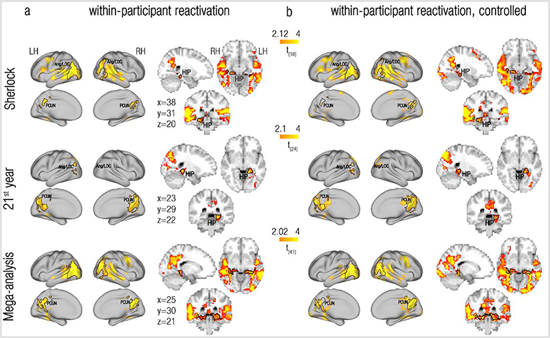
Figure 2. Within-participant reactivation of remote past events at event boundaries.
(a) Within-participant reactivation, where reactivation indices were derived from event boundary and scene representations that were extracted from the same brain. (b) Controlled within-participant reactivation, where the within-participant reactivation indices at event boundaries were contrasted with those computed for control timepoints. Rows depict within-dataset group maps of the Sherlock dataset (1st row), the 21st year dataset (2nd row) and a mega-analysis pooling together the two datasets (3rd row). Maps are presented on both flat cortical surface and on 3D slices (dataset-specific MNI coordinates are depicted). Across all datasets, reactivation of temporally-remote past events (as reflected in positive t-values) was consistently found in the bilateral precuneus/retrosplenial cortex (PCUN), the Angular gyrus/Lateral Occipital Cortex (Ang/LOC) and hippocampus (HIP). To illustrate the resemblance in results between the uncontrolled and controlled analyses, we superimposed regions of interest based on the uncontrolled within-participants analyses of each dataset ((a), marked in black contours) on the corresponding controlled reactivation maps (b). LH, left hemisphere; RH, right hemisphere. All maps were created using two-sided t-tests, and were cluster-corrected for multiple comparisons across the entire brain (p<0.005).

