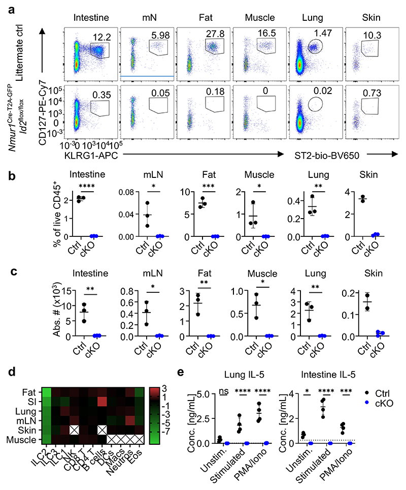
Figure 2. NmurlCre-T2A-GFP Id2flox/flox mice lack ILC2s in all tissues investigated.
a, Flow-cytometric plots of ILC2s gated from live CD45+ Lin- (CD3, CD5, CD19) cells as CD127+ KLRG1+ or CD127+ ST2+ across organs of Nmur1Cre-T2A-GFP Id2flox/floxmice (cKO) and littermate controls (Ctrl). Numbers denote percentage of cells inside the gate. b,c, Quantification of (a), Student’s t-test. d, Heatmap displaying Log2-fold changes of the median cell frequencies among live CD45+ cells across different populations and organs of Nmur1Cre-T2A-GFP Id2flox/flox mice, compared to littermate controls. X, not determined. e, IL-5 secretion measured in the supernatant of cultured immune cells isolated from lung or intestine of Nmur1Cre-T2A-GFP Id2flox/flox mice and littermate controls, stimulated with IL-2, IL-7, IL-25 and IL-33 or PMA/Ionomycin for 8 h. Each symbol represents data from one mouse, mean +/- SD, two-way ANOVA with Šídák’s multiple comparison tests, data are representative of two independent experiments with 3-4 mice per group. ns not significant, *p<0.05, ** p<0.01, *** p<0.001, **** p<0.0001. Littermate controls for a-d were Id2flox/+ and Nmur1Cre-T2A-GFP Id2flox/+combined.

