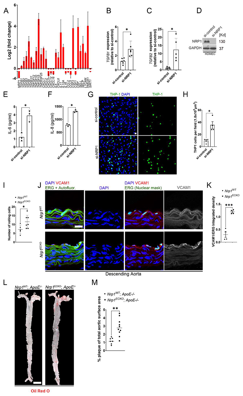
Fig. 5. NRP1 suppresses inflammatory pathways and atherosclerosis.
(A) Fold changes (Log2) in gene expression of HUVECs transfected with si-NRP1 for 72 hours and exposed to laminar flow for 24 hours relative to HUVECs transfected with si-control under the same experimental conditions. Data are presented as means ± SD. N = 3 biological replicates per group. (B) TGFB1 gene expression in HUVECs transfected for 72 hours with si-control or si-NRP1 and cultured under static conditions, expressed as fold change relative to si-control. Data are presented as means ± SD. N = 6 biological replicates per group. *p < 0.05 by paired t-test. (C) TGFB2 gene expression was measured in HUVECs transfected for 72 hours with si-control or si-NRP1 and cultured under static conditions. TGF-β2 gene expression was expressed as fold change relative to si-control. Data are presented as means ± SD. N = 5 biological replicates per group. *p < 0.05 by paired t-test. (D) Representative immunoblotting for NRP1 and GAPDH of HDMECs transfected for 72 hours with si-control or si-NRP1 and cultured under static conditions. (E and F) Quantification by ELISA of IL-6 (E) and IL-8 (F) secretion in HDMECs transfected for 72 hours with si-control or si-NRP1. Data are presented as means ± SD. N = 3 biological replicates per group. *p < 0.05 by t-test. (G) THP-1 cells labelled with Calcein-AM 1μM (green) adhering to a confluent monolayer of HUVECs transfected with si-control or si-NRP1 for 72 hours and counterstained with DAPI (blue). (H) Number of adhering THP-1 cells per field. Data are presented as means ± SEM. N = 3 biological replicates per group. *p < 0.05 by paired t-test. (I) Rolling leukocytes in the postcapillary venules of the mesentery of Nrp1WT (n=5) or Nrp1ECKO (n=7) mice injected daily with tamoxifen (12.5mg/kg) for 5 days at 4 weeks of age and imaged after 2 weeks. Data are presented as means ± SD. *p < 0.05 by unpaired t-test. (J) Rings of descending aortae from Nrp1WT or Nrp1ECKO mice injected daily with tamoxifen (12.5mg/kg) for 5 days at 4 weeks of age and immunostained after 4 weeks for ERG (green), VCAM1 (red and grey) and counterstained with DAPI (blue). Scale bar = 20 μm. ImageJ software was used to mask the autofluorescence in the green channel to reveal ERG-DAPI double positive ECs. (K) Quantification of VCAM1 integrated density relative to ERG. Data are presented as means ± SD. N = 4 mice per group. ***p < 0.001 by paired t-test. (L) En-face aortae from Nrp1WT;ApoE-/- or Nrp1ECKO;ApoE-/- mice, injected daily with tamoxifen (12.5mg/kg) for 5 days at 4 weeks and stained 18 weeks of age with Oil-Red-O (Red). Red staining shows plaque deposition on the inner wall of aortae. Scale bar = 2.1 mm (M) Quantification of plaque deposition was expressed as percentage of total aortic surface coverage. Data are presented as means ± SD. N ≥ 7 mice per group. **p < 0.005 by t-test.

