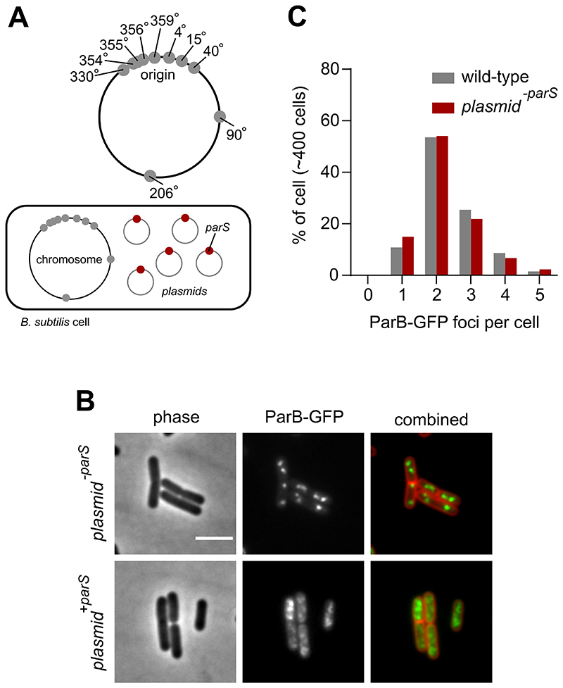
Figure 2. Extrachromosomal parS redistribute ParB.
(A) Schematic diagram showing the location of B. subtilis endogenous chromosomal parS (grey circles) and plasmid containing a single parS (red circles). (B) ParB-GFP localization in a wild-type strain harbouring plasmids that contain either a single or no parS. Phase contrast (left panel), ParB-GFP (middle panel) and membrane dye FM 5-95 combined with ParB-GFP (right panel). (C) The number of ParB-GFP foci per cell remains unchanged in the presence of the empty vector. Scale bar, 3 μm.

