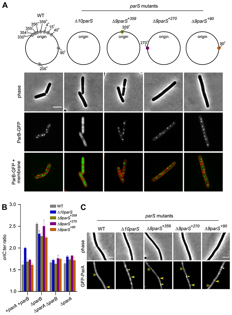
Figure 6. A single chromosomal parS is necessary and sufficient to regulate ParA activity.
(A) ParB-GFP localization in strains that differ in the location of parS. Phase contrast (top panel), ParB-GFP (middle panel) and membrane dye FM 5-95 combined with ParB-GFP (bottom panel). Schematic diagram showing the location of B. subtilis endogenous chromosomal parS (grey circles), parS at 359° (green circle), parS at 270° (purple circle) and parS at 90° (orange circle). (B) The frequency of DNA initiation was restored by ParB sliding clamps loading at a single parS. The oriC-ter ratio of each strain was determined using quantitative PCR. (C) GFP-ParA localization in strains that differ in the location of parS. A single-point arrow (->) denotes localization at a septum, a double-point arrow (-≫) indicates localization as a focus and a circle (●) denotes nucleoid binding. Scale bar, 3 μm.

