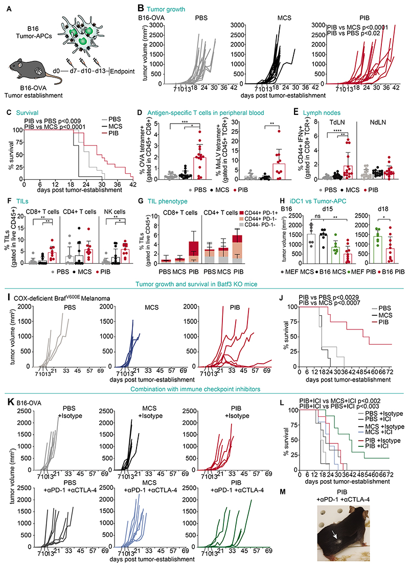
Figure 8. Tumor-APCs trigger anti-tumor immunity in vivo.
(A) B16-OVA tumors were injected intratumorally at day 7, 10 and 13 with B16-derived tumor-APCs pulsed with OVA protein and Poly(I:C). (B) Tumor growth and (C) survival of mice injected with tumor-APCs (PIB) compared with PBS and injection of control transduced cells (MCS) (n=16). (D) Flow cytometry quantification of peripheral blood T cells with OVA tetramer (left) or Murine Leukemia Virus (MuLV) tetramer (right), as a proportion of CD45+ CD8+ T cells, at day 14 after tumor establishment. (n=10-15) (E) Quantification of CD44+IFN-γ+ CD8+ T cells isolated from tumor-draining (TdLN) or non-draining lymph nodes (NdLN) after in vitro restimulation with pmel peptide at day 18. (n=15) (F) Quantification of tumor infiltrating lymphocytes (TILs) and (G) CD44+ and PD-1+ expression at day 18. (n=10) (H) Volumes of B16 tumors at day 15 (left) and 18 (right) treated with tumor-APCs (B16 PIB) compared with MEF-derived iDC1 (MEF PIB) after overnight stimulation with Poly(I:C) (n=7-10). (I) Tumor growth and (J) survival of BATF3-/- mice injected with Cox-deficient BRAFV600E melanoma tumor cells treated with tumor-APCs derived from the same cell line after overnight stimulation with Poly(I:C) (n=6-8). (K) Tumor growth and (L) survival of mice treated with ICIs (anti-PD-1 and anti-CTLA-4) or isotype controls (IgG2a and IgG2b) in combination with B16-derived tumor-APCs (PIB) after overnight incubation with OVA and Poly(I:C) (n=9-10). (M) Animal cured with combination therapy showing depigmentation (white arrow) on tumor regression site. Mean±SD is represented. n= biological replicates. P values were calculated using Kruskal Wallis (B, D-H), Mann-Whitney (H), and Mantel-Cox test (C, J, L). *P<0.05, **P<0.01, ***P<0.001, ****P<0.0001.

