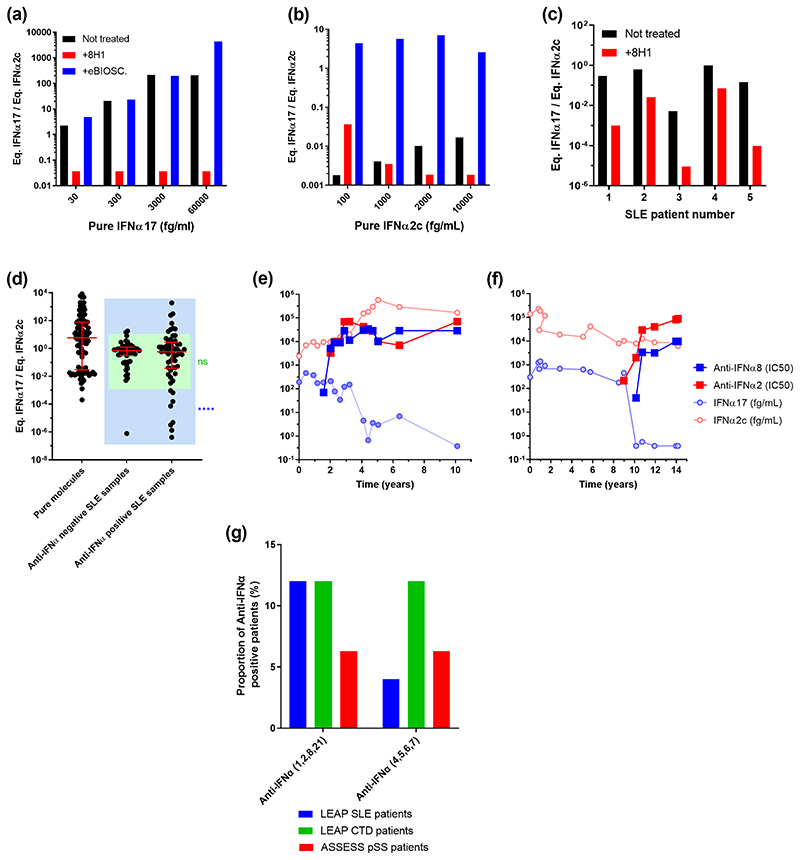
Fig. 5. Association of anti-IFNα auto-antibodies and the abnormal IFNα subtype ratios.
(a) Competition assays after addition of the 8H1 clone anti-IFNα antibody in pure IFNα17 solutions at different concentrations reduces the ratio. (b) Addition of the anti-IFNα2 antibody in pure IFNα2c solutions at different concentrations increases the ratio. (c) Addition of 8H1 in five SLE patient samples reduces the IFNα17/α2 ratio. (d) IFNα17/α2 protein ratios for SLE patient samples characterized positive for anti-IFNα antibodies in comparison with negative SLE patient samples and pure molecules. (e-f) Longitudinal analysis of anti-IFNα8 and anti-IFNα2 antibody concentrations, pan-IFNα and IFNα2 assays results over time (years) in two SLE patients. (g) Proportion of anti-IFNα auto-antibodies positive samples in LEAP SLE, LEAP CTD and ASSESS pSS patients.

