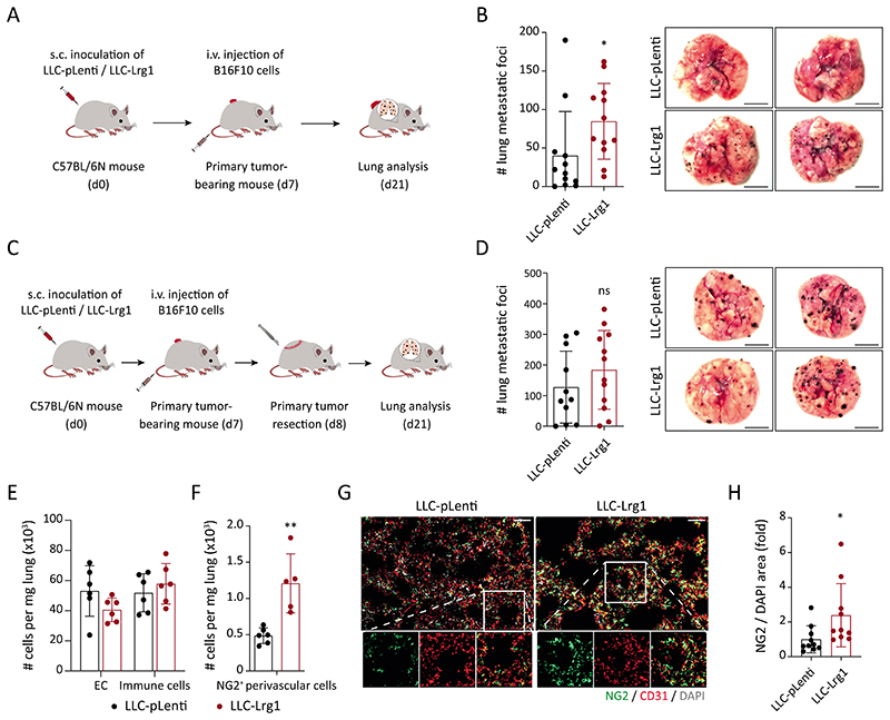
Fig. 5. Systemic upregulation of LRG1 promotes metastasis.
(A) Lrg1-overexpressing LLC (LLC-Lrg1) or control-LLC (LLC-pLenti) cells were subcutaneously inoculated in C57BL/6N mice. 7 days after inoculation, melanoma (B16F10) cells were intravenously injected to initiate an experimental metastasis assay. (B) On the left, dot plot showing the number of melanoma metastases in the lung, and on the right, representative lung images (mean ± SD, n = 12 mice). Scale bars = 5 mm. *, P<0.05 (two-tailed Mann-Whitney U test, comparing to LLC-pLenti). (C) C57BL/6N mice were injected subcutaneously with LLC (pLenti/Lrg1) cells. On day 7, tumor-bearing mice were injected intravenously with melanoma (B16F10) cells. Primary tumors (the source of LRG1) were resected 24 hours after the intravenous injection of melanoma cells. (D) On the left, dot plot showing the number of melanoma metastases in the lung, and on the right, representative lung images (mean ± SD, n = 11-12 mice). The comparison was rendered non-significant (ns, P= 0.31) according to two-tailed Mann-Whitney U test. (E-H) WT or NG2-Cre X YFPfl/fl mice were injected with either LLC-Lrg1 or LLC-pLenti cells. FACS-based quantitation of ECs, immune cells, and NG2+ perivascular cells in the lung of tumor-bearing mice (E, F) (mean ± SD, n = 5-6 mice). **, P<0.01 (two-tailed Mann-Whitney U test, comparing to LLC-pLenti). Lung tissue sections were stained for NG2 (pericyte-specific). Representative images of lung sections (G). Scale bars = 50 μm. Quantitation of NG2/DAPI area is shown (H) (mean ± SD, n = 10 mice). *, P<0.05 (two-tailed Mann-Whitney U test, comparing to LLC-pLenti).

