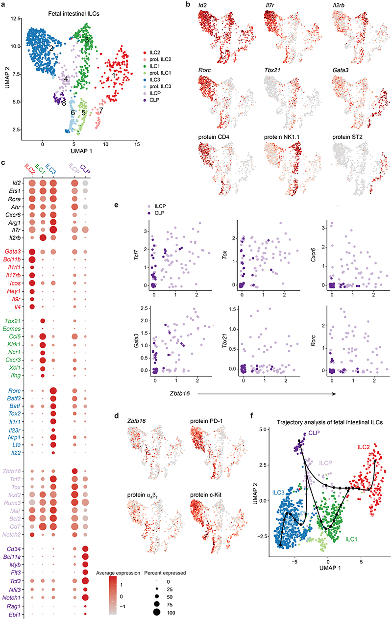
Fig. 1. Single-cell sequencing of fetal intestinal cells reveals a spectrum of mature ILC subsets and progenitors.
Viable Lin(CD19, CD3, CD5, F4/80, FcɛRIα, Gr-1)- CD45+ cells expressing IL-7 receptor (CD127) and/or the IL-2 receptor subunit beta (CD122) isolated from the small intestine (SI) of E18.5 Rorc(gt)GFP/wt embryos were sort-purified by flow cytometry, and a single-cell expression library was generated using 10x Genomics. a, Uniform Manifold Approximation and Projection (UMAP) identifies eight distinct clusters. (b) Gene expression and Cellular Indexing of Transcriptomes and Epitopes by Sequencing (CITE-seq) protein expression UMAP plots. (c) Selected gene expression within clusters. Colour scale represents average expression, dot size visualizes fraction of cells within the cluster expressing the gene. (d) Selected gene expression and CITE-seq protein expression UMAP plots. (e) Zbtb16 co-expression plots of selected genes within CLP and ILCP cluster. (f) Trajectory analysis using Slingshot. Inferred trajectories are represented as lines starting within the CLP cluster. Dots represent knots.

