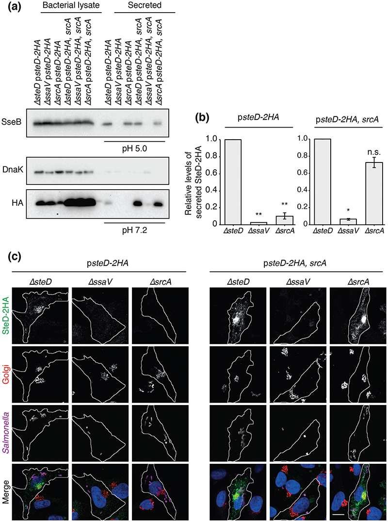
Fig. 2. SrcA is required for secretion and translocation of SteD into host cells.
(a) The indicated Salmonella strains were grown in minimal medium pH 5.0 for 4h then washed and transferred to minimal medium pH 7.2 for 2 h. All Salmonella strains carried plasmid pWSK29 expressing SteD-2HA under its endogenous promoter, with or without SrcA. Bacterial lysate proteins and secreted proteins at either pH 5.0 or pH 7.2 were examined by immunoblotting with antibodies against DnaK, the HA epitope or SseB. (b) Levels of secreted SteD-2HA were calculated by densitometry from immunoblots against HA using Image Lab software. Secreted protein levels from the ΔssaV and ΔsrcA strains are shown in relation to the ΔsteD strain. Mean of three independent experiments ±SD. The log10 of the ratios of secreted protein levels from the ΔssaV or ΔsrcA over that of the ΔsteD strain were analysed by one sample t-test, **P<0.01, *P<0.05. (c) Representative confocal immunofluorescence microscopy images of SteD-HA localization in Mel Juso cells at 20 h p.i. with the indicated Salmonella strains. Cells were fixed and processed for immunofluorescence microscopy by labelling for SteD-2HA (anti-HA antibody, green), the Golgi network (anti-GM130 antibody, red), Salmonella cells (anti-CSA-1 antibody, magenta) and DNA (DAPI, blue). Scale bar, -10 μm.

