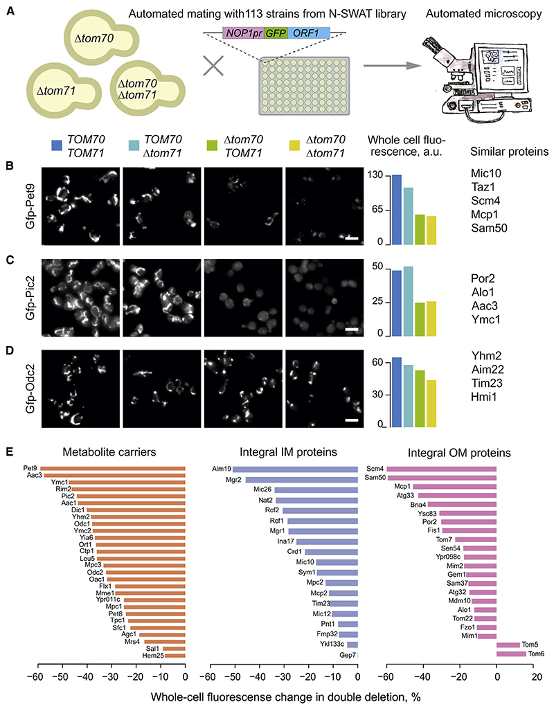
Figure 2. Mitochondrial proteins strongly differ in their Tom70 dependence.
(A) Scheme of the systematic visual screen of GFP-tagged mitochondrial proteins.
(B–D) The mitochondrial localization of 113 N-terminally GFP-tagged mitochondrial proteins (all lacking an MTS) were visualized. Proteins shown in (B) showed a strongly reduced mitochondrial localization in the absence of Tom70 and moderately reduced levels if Tom71 was deleted. Thus, these proteins depend to some degree on both receptors. Proteins shown in (C) were unaffected if Tom71 was deleted but still required Tom70. For proteins shown in (D), Tom70 and Tom71 were hardly, if at all, relevant.
(E) The whole-cell GFP signal change in Δtom70Δtom71 compared with wild-type cells measured for different mitochondrial protein classes. See Table S3 for details. Scale bars, 10 μm. OM, outer membrane.

