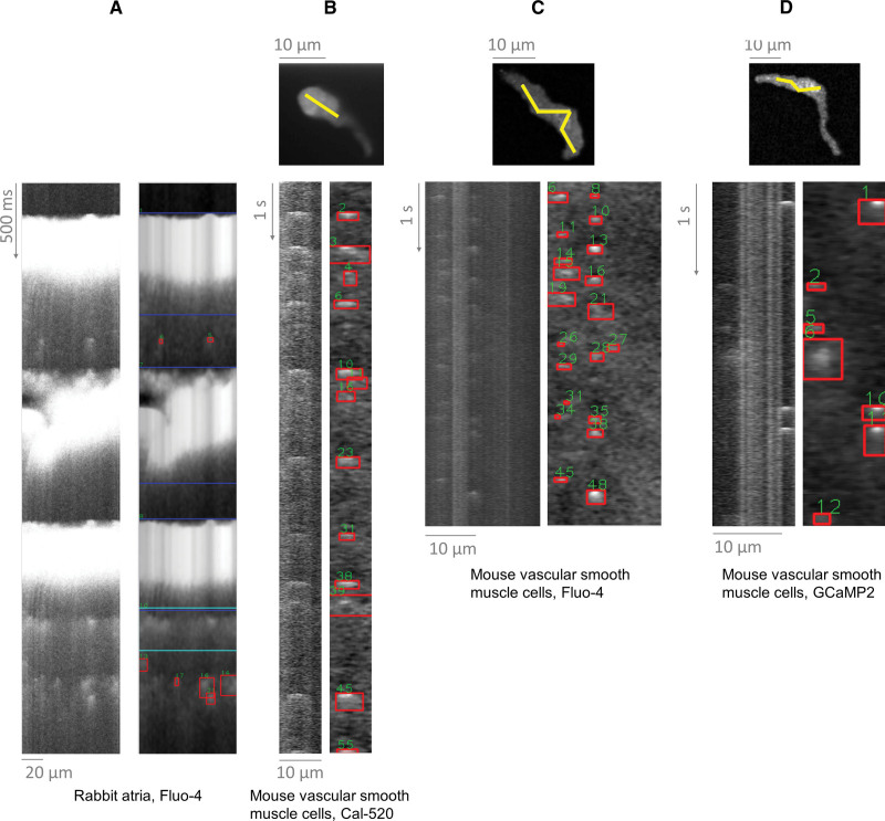
Figure 6.
Application of SparkMaster 2 (SM2) to distinct cell types and conditions. A, Paced rabbit atrial cells loaded with Fluo-4. B, Mouse vascular smooth muscle cells imaged using a spinning disk confocal microscope and the Ca dye of Cal-520. Top image shows recorded images, with yellow line being used to extract the line-scan recording underneath. C and D, Analogous recordings of vascular smooth muscle cells, using Fluo-4 and GcaMP2, respectively to image the Ca sparks. Performance of the original SparkMaster on these recordings is shown and discussed in Supplemental Material S3.

