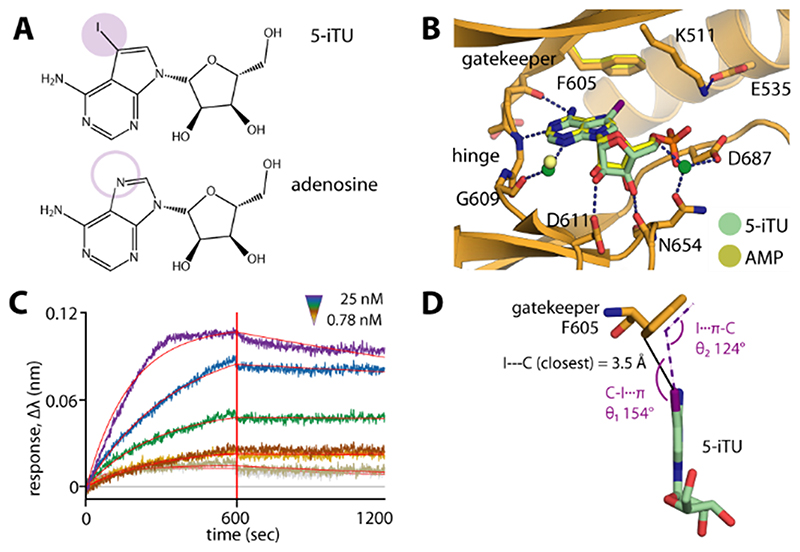
Figure 1. The 5-iodotubercidin inhibitor (5-iTU) exhibits tight binding with slow dissociation kinetics from haspin.
A) Chemical structures of 5-iTU and adenosine. B) Superimposition of haspin–5-iTU and AMP (PDB ID: 4ouc) reveals similar binding modes for the two compounds. C) The BLI sensorgram suggests slow kinetics for the 5-iTU–haspin interaction. D) The iodide and benzene moieties of 5-iTU and F605, respectively, are located in close proximity, with a favorable geometry for a halogen–π bond.

