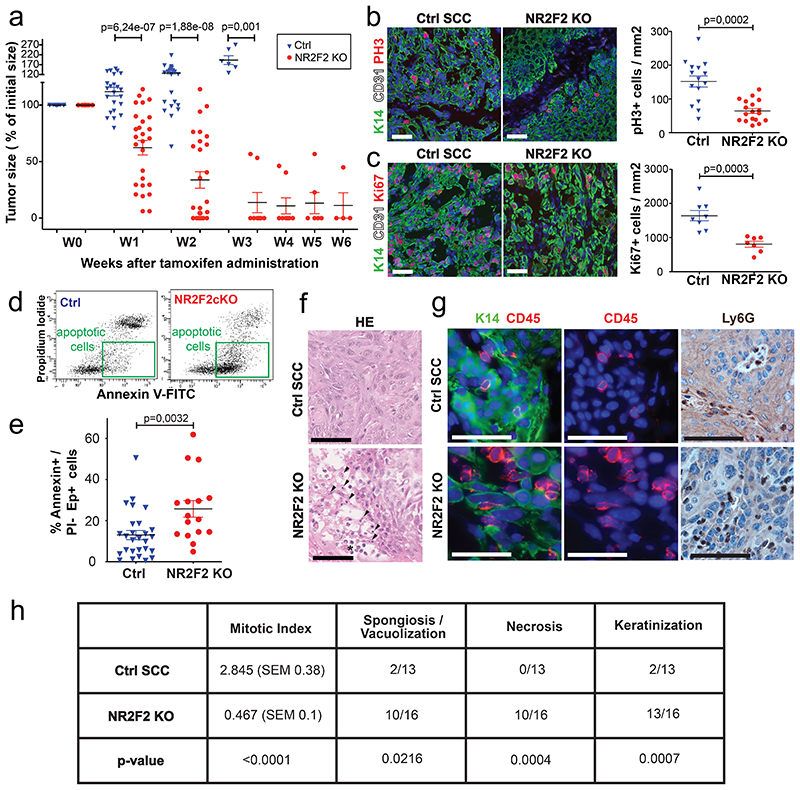
Figure 4. NR2F2 is essential for the maintenance of malignant SCCs.
a: Percentage of initial tumor size over time following tamoxifen induced NR2F2 deletion (n=21 Ctrl and 28 NR2F2 KO tumors).
b: Immunostaining (left) for pH3 (mitotic cells) and K14 (Tumor cells) and quantification (right) of pH3+ cells/mm2 (n=15 Ctrl and 18 NR2F2 KO tumors).
c: Immunostaining (left) for Ki67 (proliferating cells) and K14 (Tumor cells) and quantification (right) of Ki67+ cells/mm2 (n=8 Ctrl and 7 NR2F2 KO tumors).
d: FACS plot of tumor cells stained with AnnexinV and PI. e: Quantification of AnnexinV+/PI- Epcam+ cells (early cell death) in Ctrl and NR2F2 KO tumors (n=27 Ctrl and 16 NR2F2 KO tumors). f: HE of Ctrl and NR2F2 KO SCC. NR2F2 KO tumors display necrotic areas with picnotic nuclei (arrowheads) and polymorphonuclear cells recruitment (asterisk).
g: Immunostaining for CD45 (immune cells) and K14 (tumor cells) and IHC for Ly6G (neutrophils) showing immune cell infiltration of the tumor following NR2F2 deletion. Representative images of at least 5 independent biological replicates per condition.
h: Summary table of the histopathological analysis of Ctrl and NR2F2 KO tumors, considering mitotic index (per high-power field), necrosis, spongiosis/vacuolisation and keratinization. (n=13 Ctrl and 16 NR2F2 KO tumors).
Scale bar = 50μm. Data in a, b, c, e are represented as mean ± SEM. The p-values are calculated using a two-tailed Mann-Whitney test. For the table h, we used a two-tailed Mann-Whitney test for the mitotic index, two-tailed Fisher’s exact test in all other cases.

