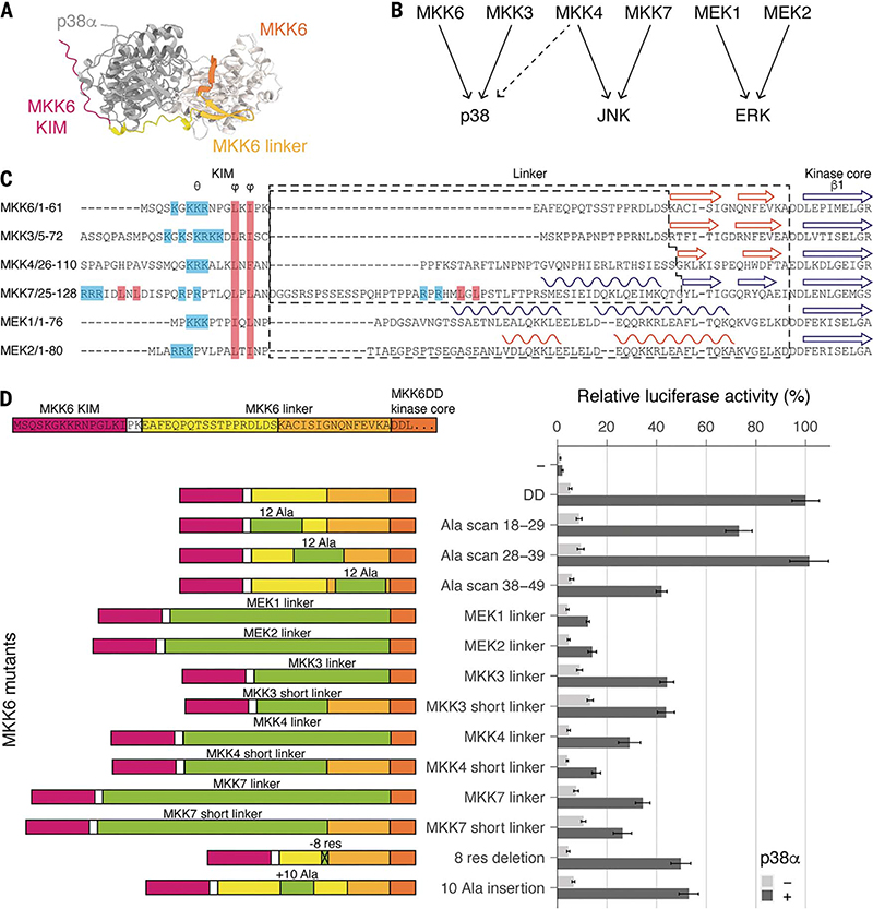
Fig. 5. MKK6 N-terminal extension length and secondary structure define the specificity of p38α activation.
(A) Top view of an MKK6-p38α AlphaFold2 multimer model, for illustrative purposes. (B) Specificity of the MAP2K/MAPK signaling pathways. (C) Sequence alignment of MAP2K N termini. Secondary structure elements are indicated, from experimental structures in blue and AlphaFold2 predictions [predicted local difference distance test (pLDDT) score > 65] in orange. (D) Luciferase reporter assay to monitor the activity of the p38α signaling pathway in HEK293T cells, showing the ability of MKK6DD mutants and chimeras to activate p38α (statistical analysis in table S9 and protein expression levels in fig. S10).

