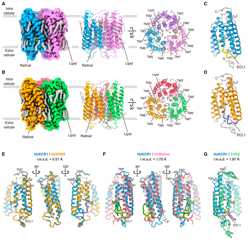
Figure 1. Cryo-EM structures of WT HcKCR1 and HcKCR2.
(A) Cryo-EM density map (left) and ribbon representation of HcKCR1 homotrimer viewed parallel to membrane (middle) and from intracellular side (right).
(B) Cryo-EM density map (left) and ribbon representation of HcKCR2 homotrimer viewed parallel to membrane (middle) and from intracellular side (right).
(C and D) Monomeric structures of HcKCR1 (C) and HcKCR2 (D).
(E–G) Structural comparisons of HcKCR1 (blue) with HcKCR2 (orange) (E), ChRmine (red) (F), and C1C2 (green) (G) from different angles.

