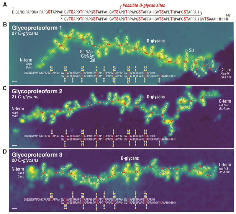
Fig. 4. STM imaging of single O-glycoproteins.
(A) Sequence of MUC1 reporter (148aa) containing 6.5 tandem repeats of 20 amino acids (GVTSAPDTRPAPGSTAPPAH) decorated with O-glycans at the Ser (S) and Thr (T) residues (total of 34 potential O-glycosites). Imaging single MUC1 proteins reveals the number, the structure, and the attachment site of O-glycans decorating the protein, as shown for a glycoproteoform with 27 O-glycans in (B), 21 O-glycans in (C), and 20 O-glycans in (D). The positions of S and T residues are indicated by red dots along the protein backbone (white dashed line). The unannotated STM images are given in Fig. S8. Scale bar is 1 nm.

