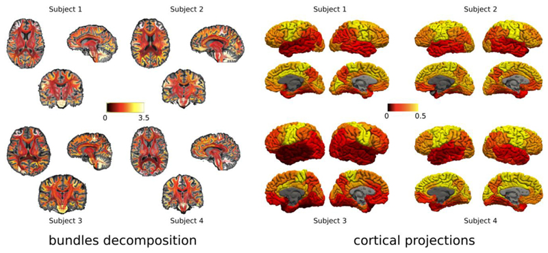
Fig. 6.
Consistency of our method across the four different subjects who were scanned with the same acquisition protocol. For each subject, on the left we show the bundles color-coded by the obtained bundle myelin fraction obtained from MySD, while on the right we present the corresponding cortical projection. In both cases the contrast between different bundles and cortical regions obtained using the MySD approach on MWF maps are highly consistent across all the subjects with local differences comparable with intrinsic differences in MWF values in the white matter of each subject. Color bars are without units because they refer to fractions of myelin and we used the same interval of values for all the subjects.

