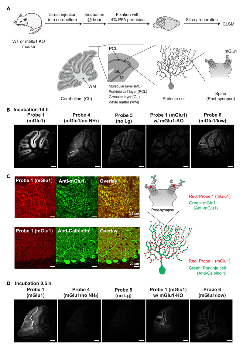
Figure 3. FixEL of a small molecule ligand for mGlu1 in the mouse cerebellum.
(A) Schematic illustration of experimental procedures. PBS(–) containing probe 1 (mGlu1) (4.5 μL of 10 μM probe which is assumed to be ca. 0.7 μM (Ccere) on the basis of the cerebellum volume33) was injected into mouse cerebellum. After 0.5 or 14 h of incubation, the mouse was transcardially perfused with 4% PFA. The cerebellum was isolated and sectioned by cryostat (50-μm thick).
(B) Fluorescence imaging of cerebellum slices with FixEL of probe 1 (mGlu1) after 14 h incubation. Imaging was performed using a CLSM equipped with a 10× objective and a GaAsP detector (633 nm excitation for Ax647). From left to right images, probes 1 (mGlu1), 4 (mGlu1/no NH2), 5 (no Lg), and probe 1 (mGlu1) in mGlu1 KO mouse, and probe 6 (mGlu1/low) were used. Scale bars, 500 μm.
(C) Co-immunostaining of the cerebellum slice with FixEL of probe 1 (mGlu1) after 12 h incubation. Probe 1 (mGlu1) is shown in red. Anti-mGlu1 and anti-calbindin are shown in green. The slices after FixEL of probe 1 (mGlu1) were permeabilized and immunostained with primary antibody anti-mGlu1 or anti-calbindin and secondary antibody (Alexa Fluor 488 [Ax488]) in 0.1% Triton X-100/PBS(–). Fluorescence images in top and bottom were acquired by using a CLSM equipped with a 100× objective, a GaAsP detector (488 nm excitation for Ax488 and 633 nm excitation for Ax647), and lightning deconvolution. Fluorescence image in middle was acquired by using a CLSM equipped with a 63× objective and a GaAsP detector (488 nm excitation for Ax488 and 633 nm excitation for Ax647).
(D) Fluorescence imaging of cerebellum slices with FixEL of probe 1 (mGlu1) after 0.5 h incubation. Imaging was performed using a CLSM equipped with a 5× objective. Scale bars, 500 μm.
See also Figure S3.

