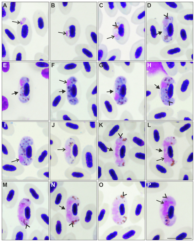
Fig. 2.
Gametocytes of Haemoproteus dumbbellus n. sp. (lineage hEMCIR01) from the blood of a yellowhammer Emberiza citrinella. Developmental stages are (A-C) young gametocytes; (D-I) macrogametocytes; (J-P) microgametocytes. The following forms can be distinguished among them: (D, E) growing macrogametocytes, (F, G) advanced macrogametocytes, (H, I) fully grown macrogametocytes, (J, K) growing microgametoccytes, (L-M) advanced microgametocytes, and (N-P) fully grown microgametocytes. Note that early gametocytes (A) do not adhere to the erythrocyte nuclei, but all other blood stages (B-P) adhere to them. The long arrow indicates the parasite nucleus and the arrowheads indicate pigment granules. An unfilled space (indicated by the short arrow in D-H, K, L, N) is present between gametocytes and the envelope of erythrocytes from the stage of developing gametocytes to the stage of fully grown gametocytes. This gives the parasite a dumbbell-like shape at most stages of growth, which is a characteristic feature of this parasite species. Note that this space often maintains in fully grown gametocytes (H, N), a rare character in Haemoproteus species. Fully grown gametocytes fill erythrocytes till the poles; they enclose the nuclei of erythrocytes with their ends but do not encircle them completely (H, I, O, P). The macrogametocyte nucleus is subterminal in position; it does not adhere to the erythrocyte nucleus (F-I). Scale-bar = 10 μm.

