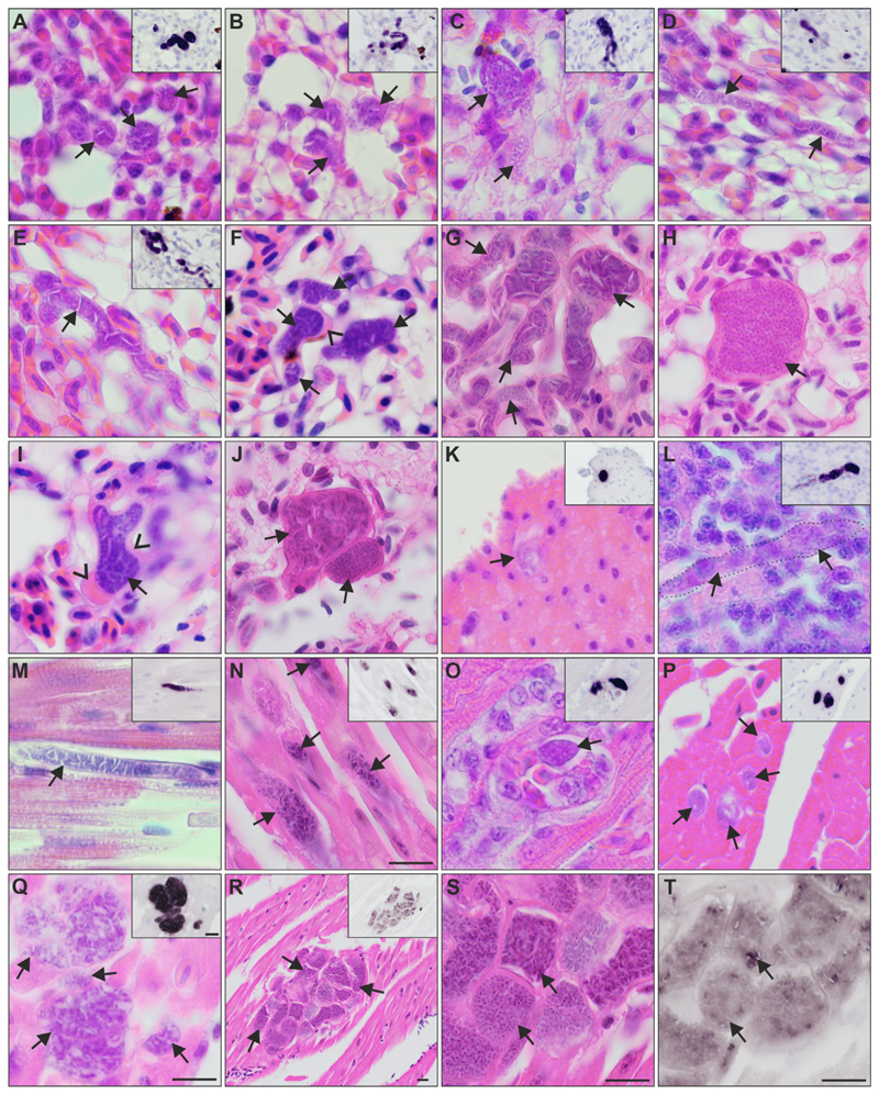
Fig. 4.
Meronts of Haemoproteus dumbbellus (lineage hEMCIR01) found in (A-J) the lungs, (K) gizzard, (L) brain, and (M-T) heart of two yellow hammers Emberiza citrinella. The meront generic origin in H & E-stained preparations was confirmed by chromogenic in situ hybridization (CISH) using a Haemoproteus genus-specific probe indicated by a purple signal in the insets of panels A-E and K-R. Independently of their maturation stage, meronts (indicated by arrows) differed greatly in shape, being round (A, B, K), oval (O, P) or elongate (D, L-N), and ranging in size from less than 10 μm (P) to more than 50 μm (D, E, M, Q). Elongate meronts in the heart appear to follow the shape of the muscle cells (M, N), whereas meronts in the lung tissue (E) were often of capillary shape. Meronts were surrounded by a thin eosinophilic wall, occasionally with bulges of unclear origin located at the periphery of nearly mature parasites (F, I). Note the development of angular-shaped cytomeres separated by clefts, which is a characteristic feature of meront maturation in this parasite species. Such clefts are particularly well visible between cytomeres before merozoite formation (E, G, J-upper arrow), and they disappear in mature meronts, which are overfilled with discrete merozoites (H, J-lower arrow). R-T show the same group of meronts at different magnifications. Note the difference in maturation among meronts (R-T), with mature, roundish merozoites, characterized by weak CISH signals (S, T, lower arrow), or the still developing cytomeres and stronger CISH signals (S, T, upper arrow). Arrowheads indicate the eosinophilic wall. Scale-bar = 10 μm.

