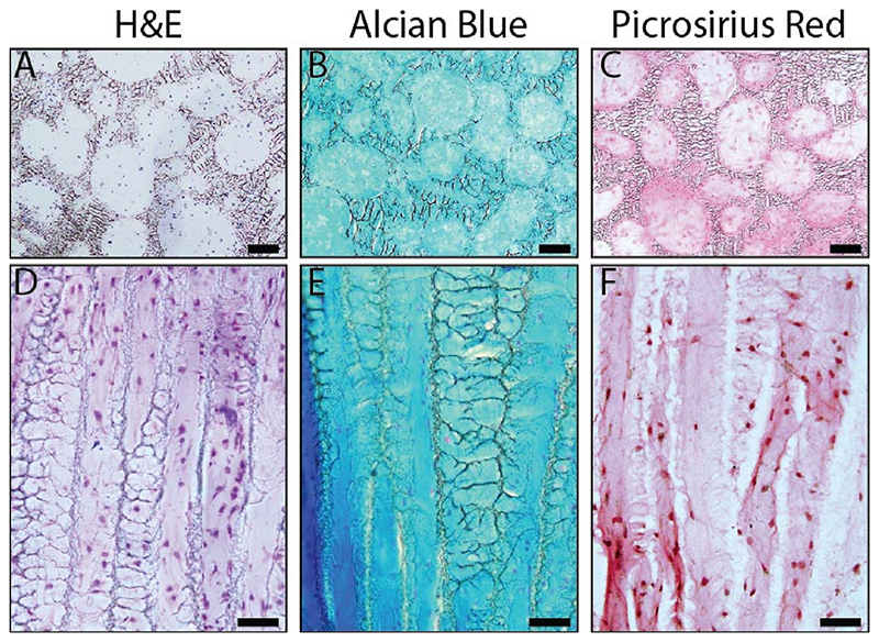
Fig. 3.
Histological analysis of tissue formed within the scaffold, seeded with 10 × 106 bovine chondrocytes, and cultured in vitro under chondrogenic conditions for 4 weeks. The PLZ (A-C; scale bars = 100 μm) and DFZ (D-F; scale bars = 50 μm) were stained with (A,D) hematoxylin and eosin (H&E) for nuclear morphology and non-specific tissue visualization, (B,E) Alcian blue for GAG, and (C,F) picrosirius red for collagen deposition. (For interpretation of the references to color in this figure legend, the reader is referred to the Web version of this article.)

