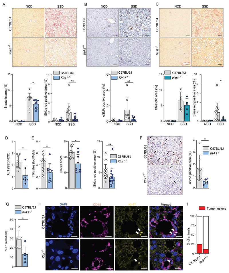
Figure 3. NKG2D engagement is essential for development of liver fibrosis in context of NASH.
(a-b) WT and Klrk1-/- (NKG2D-deficient) mice were fed an NCD or an SSD diet for 16 weeks (n=4-11). (a) (top) Representative liver slides stained with Sirius Red (200 X) and (bottom) quantification of steatosis and fibrosis. Scale bars indicate 100 µm. (b) (top) Representative liver slides stained for αSMA and (bottom) quantification of hepatic stellate cell activation (200 X). Scale bars indicate 100 µm. (c) WT and Hcst-/- (DAP10-deficient) were fed an NCD or an SSD diet for 16 weeks. (top) Representative liver slides stained with Sirius Red (200 X) and (bottom) quantification of steatosis and fibrosis by Sirius red staining Scale bars indicate 100 µm. (n = 4-5). (d-i) WT and Klrk1-/- mice were fed an NCD or an SSD diet for 52 weeks and livers were analyzed (n=7-9). (d) quantification of ALT in serum. (e) quantification of liver pathology of histological slides stained with H&E or Sirius red. (f) (left) Representative liver slides stained for αSMA and (right) quantification of hepatic stellate cell activation (200 X). Scale bars indicate 100 µm. (g) slides were stained for Ki67 and positive cells per field of view were quantified. (h) Representative immunofluorescence staining of CD45 and Ki67. Arrows mark Ki67+ nuclei. Scale bars indicate 10 µm. (i) number of mice carrying macroscopically visible tumors were quantified (n=17-20). The data are representative of at least two independent experiments or show pooled data of two experiments. Shown are means +/- s.e.m. Statistical significance was determined by unpaired t test. Statistical significance was defined as *p<0.05; **p<0.01; ***p<0.001.

