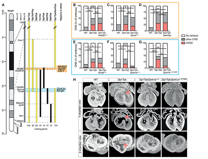
Figure 5. Three copies of Dyrk1a are necessary to cause heart defects.
(A) Map of Hsa21 showing regions of orthology to Mmu10, Mmu17 and Mmu16 (grey) and indicating regions of Mmu16 that are duplicated in mouse strains that show CHD (yellow) and in those that do not (black); genes at boundaries of these duplications are indicated next to the Mmu16 map; numbers of coding genes indicated below duplicated regions. Two genetic intervals containing 3 and 2 candidate genes for CHD are indicated in orange and blue, respectively. (B-G) Graphs of percentage of CHD in E14.5 mouse embryonic hearts from the indicated mouse models, indicating the frequency of AVSD and other CHD, which are predominantly VSDs and occasionally outflow tract defects such as overriding aorta. The orange and blue boxes highlight the genes found in the 2 candidate regions. The cross of Dp1Tyb to Del4Tyb was used to determine if 3 copies of Cbr1 are required for heart defects. Numbers of embryonic hearts analyzed: (B) WT (n=25), Dp1Tyb (n=20), Dp1TybMir802+/+/- (n=27) hearts with two copies of Mir802; (C) WT (n=24), Dp1Tyb (n=23), Dp1TybSetd4+/+/- (n=26) hearts with two copies of Setd4; (D) WT (n=7), Dp1Tyb (n=13), Dp1TybDel4Tyb (n=15) hearts with two copies of the region deleted in Del4Tyb; (E) WT (n=34), Dp1Tyb (n=41), Dp1TybKcnj6+/+/- (n=30) hearts with two copies of Kcnj6; (F) WT (n=91), Dp1Tyb (n=86), Dp1TybDyrk1a+/+/- (n=23) hearts with two copies of Dyrk1a; (G) WT (n=30), Dp1Tyb (n=23), Dp1TybDyrk1a+/+/K188R (n=44) hearts with two WT and one kinase-inactive allele of Dyrk1a. Fisher's exact test, * 0.01 < P < 0.05, ** 0.001 < P < 0.01, *** P < 0.001 for difference in number of total CHD, except for Dp1TybDyrk1a+/+/K188R cohort, where statistics were calculated for number of AVSD; ns, not significant. (H) 3 Dimensional high resolution episcopic microscopy (HREM) rendering of WT, Dp1Tyb, Dp1TybDyrk1a+/+/- and Dp1TybDyrk1a+/+/K188R mouse hearts, eroded to show an anterior four-chamber view (top and middle) and a two-chamber view (bottom) seen from the atria at the level of the atrioventricular canal. Top and bottom rows show eroded 3D views, middle row shows 2D sections at the same level as shown in the top row. Red arrowheads indicate VSD (top, middle) or AVSD (bottom). iAVC, inferior atrio-ventricular cushion; LV, left ventricle; MV, mitral valve; RV, right ventricle; sAVC, superior atrio-ventricular cushion; TV, tricuspid valve; VS, ventricular septum.

