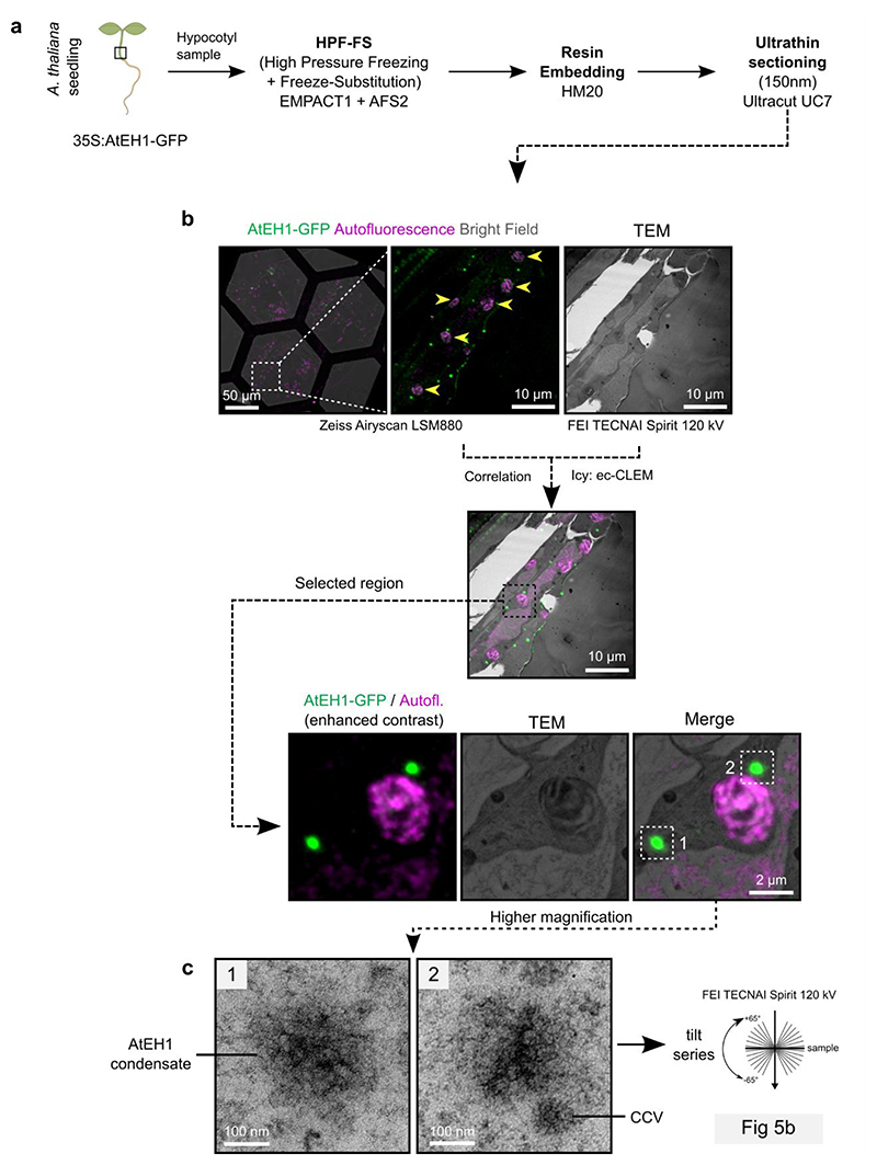
Extended Data Fig. 5 (Related to Figure 5). CLEM-ET workflow.
a, Overview of the sample preparation procedure. b, Expanded workflow of the Correlative Light and Electron Microscopy (CLEM) imaging of hypocotyl sections. Yellow arrowheads indicate chloroplasts used as natural landmarks for correlation. c, Zoomed images of regions selected for electron tomography (ET) reconstruction. The condensates have similar electron density to CCVs.

