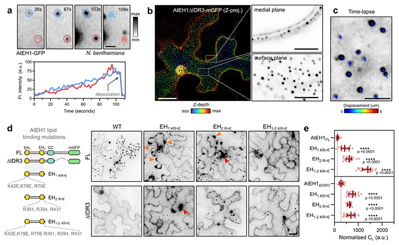
Figure 3. AtEH1 condensates nucleate on the plasma membrane via phospholipid binding domains.
a, Time-lapse imaging of UBQ10:AtEH1-GFP in N. benthamiana epidermal cells. Condensates appear as immobile plasma membrane associated puncta that gradually increase in intensity (blue and red circles), before dissociating from their original location (arrows). b, Depth colour-coded projections of UBQ10:AtEH1ΔIDR3-mGFP in N. benthamiana epidermal cells. Insets (single Z-plane sections) show that the condensates are restricted to the plasma membrane. c, Tracking of condensate displacement over 1 minute. d, Schematic of EH domain lipid binding mutants in AtEH1FL and AtEH1ΔIDR3 constructs and representative confocal images of UBQ10:AtEH1-GFP reporter constructs expressed in N. benthamiana epidermal cells. Orange arrowheads indicate irregular condensates appearing at cell lobes, red arrowheads indicate condensates with abnormal morphology. Condensates were not observed in AtEH1FL when both EH domains were mutated. e, Quantification of light phase (CL). Data is median ± 95% CI. Statistics indicates significance to the control (AtEH1FL or AtEH1ΔIDR3), **** = p <0.0001, unpaired t-test with Welch’s correction; n = 18, 16, 18, 20, 17, 18, 16, 21 cells. Scale bars = 2 μm (a), 5 μm (b; inset), 20 μm (c, d). See also Extended Data Fig. 3; Video S1. Experiments were performed three times with similar results. Source data are provided as a Source Data file.

