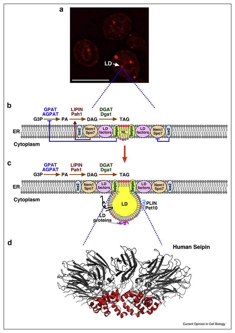
Figure 2. The ordered formation of LDs at discrete sites in the ER membrane.
a) LD biogenesis occurs at discrete ER domains. Fluorescence microscopy image of a yeast strain expressing mCherry-tagged Seipin, induced to form TAG, which drives de novo formation of LDs. Scale bar, 5 μm. b, c) Schematic view of interactions between components needed for LD formation. Seipin is required at the center to promote NL nucleation within the ER membrane. Seipin is assisted by LD factors such as Pex30, and LDAF1/Promethin (see Table 1), and controls the production of PA. The Nem1/Spo7 complex activates Lipin to promote DAG formation, which then serves as substrate for TAG synthesis by DGAT enzymes. Nem1/Spo7 activity is inhibited by Ice2. Upon LD growth and maturation, LD proteins including perilipins (PLIN) localize onto the limiting monolayer. DAG in the ER membrane is depicted by red circles and TAG by the yellow sphere. d) Model of the oligomeric structure of human Seipin. NL nucleation is promoted by the membrane apposed hydrophobic helix (red) within the luminal domain of the Seipin inner ring. The two transmembrane domains of Seipin are not shown.

