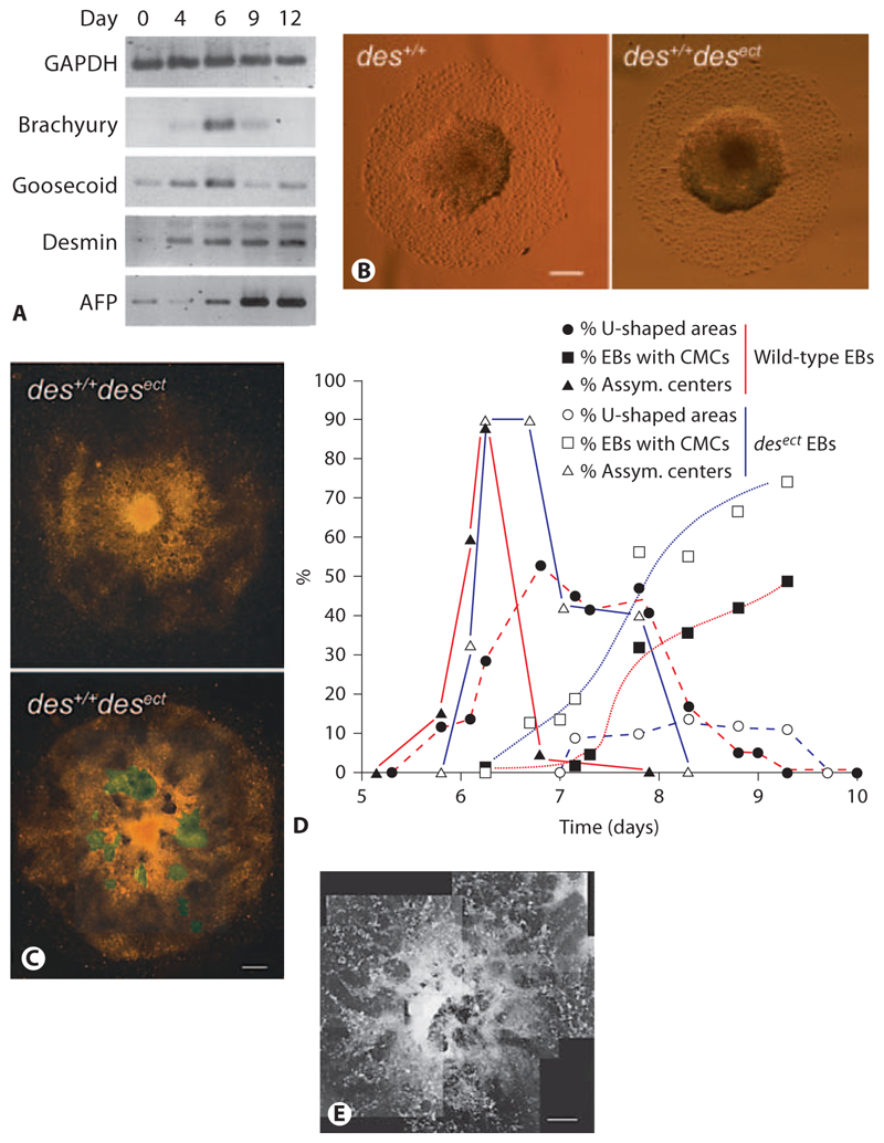
Fig. 6. Ectopic expression of the early muscle-specific protein Desmin disturbs morphogenesis in EBs.
A Semiquantitative RT-PCR analysis of mRNAs isolated from developing EBs at times indicated. GAPDH was loading control. Alpha fetoprotein (AFP) was used as control to monitor differentiation of endoderm. B Typical morphology of wild-type (des+/+) and des+/+ desect EBs at days 6–7. C des+/+desect EBs at days 8–9. D Development of asymmetric centers in EBs (▲), horseshoe-shaped ridges surrounding the centers (●), and rhythmically contracting CMCs (◼) in wild-type (red lines; n = 788) and des+/+ desect (blue lines; n = 706) EBs. Standard deviation was always less than 15% and is omitted for clarity reasons. E An aggregate of somatic stem cells isolated from murine hearts at day 13 after aggregation. B Phase-contrast images. C, E Dark-field pictures composed of several images. Areas with rhythmically contracting CMCs are highlighted in green. Bars: 200 μm (B), 100 μm (C), 1 mm (E).

