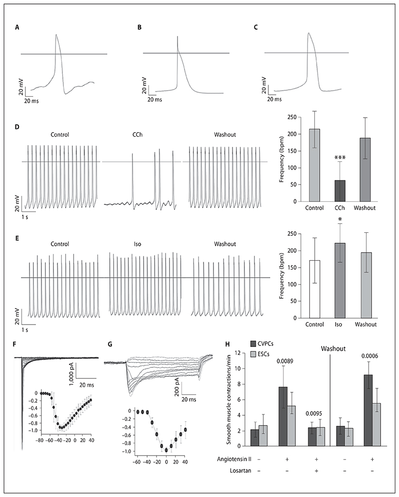
Fig. 5. Electrophysiological characteristics of CVPC-derived CMCs and angiotensin II dependent contraction of SMCs.
A–C CMCs with pacemaker-, atrial-, or ventricular-like action potentials. Effects of muscarinic (D) and β-adrenergic (E) stimulations of CMCs. Negative chronotropic effect of 1 μmol/l carbachol (CCh) (D) and positive chronotropic effect of 1 μmol/l isoproterenol (Iso) (E). Representative experiments (left), and impact on beating frequency (right). Number of cells used for the analysis: carbachol, 14; isoproterenol, 12. * p < 0.05, *** p < 0.001. Functional expression of cardiac voltage-gated sodium channels (F) and L-type Ca2+ channels (G). Insets: current amplitudes normalized to their maxima and plotted against test voltages. H Angiotensin II increases the contraction rates of SMCs. CVPCs and ESCs were aggregated and SMCs were monitored for spontaneous contractions for 10 min on days 21 and 22 after aggregation. Then 1 μmol/l angiotensin II was added to the plates and contraction was again monitored for 10 min. After that 1 μmol/l losartan was added to block the angiotensin II receptor and measurements were repeated. After washout of both angiotensin II and losartan, CBs and EBs were incubated for 30 min at 37°C and then 1 μmol/l angiotensin II was again added. Number of experiments = 5 with 2 CVPC lines. Error bars indicate the standard deviation. p values are given for CVPC-derived SMC contraction rates in relation to the temporally preceding values.

