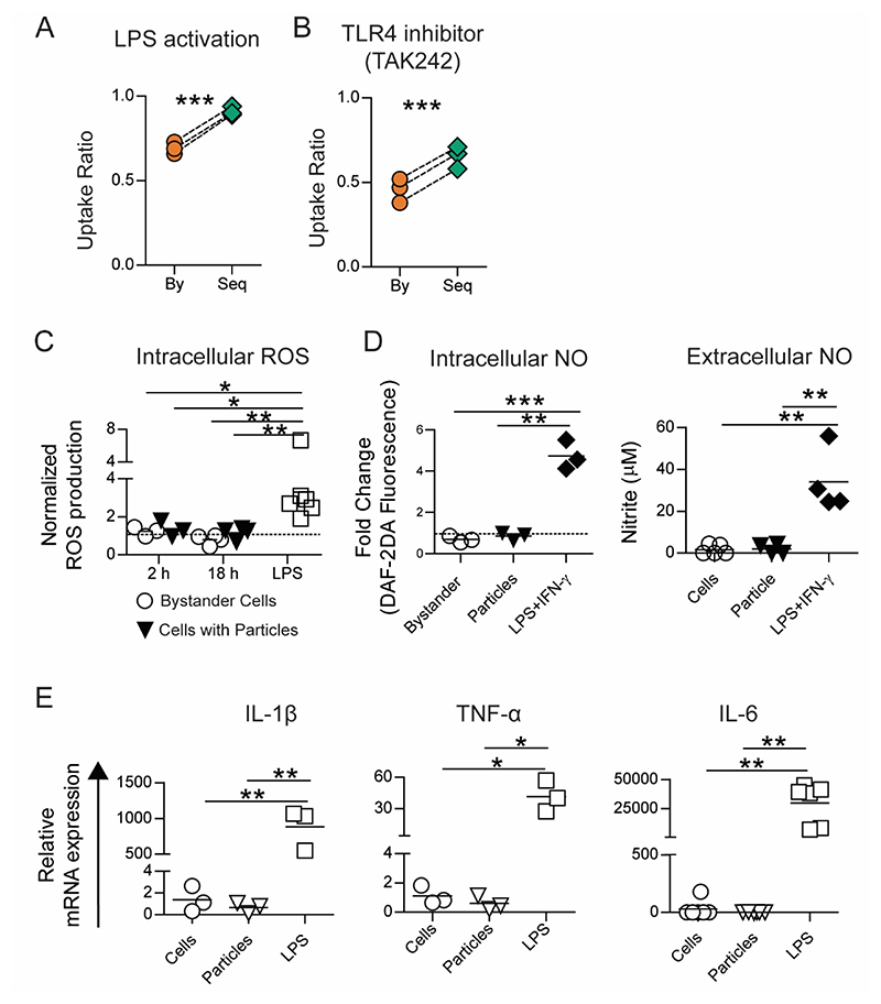
Figure 3. Sequential uptake is not due to TLR based cellular activation.
A − Uptake of 500 nm-carboxylated polystyrene (PS) particles by RAW macrophages (1:50 cell to particle ratio) following pre-activation of all cells with LPS (1 μg/ml for 18 hours). B − Sequential uptake of 500 nm-carboxylated PS particles by RAW macrophages following the treatment of cells with TAK-242 (2 μM for 6 hours) to block TLR-4 mediated cellular activation. C − Intracellular reactive oxygen species (ROS) quantified using flow cytometry following incubation of RAW macrophages with 500 nm-carboxylated PS particles (1:50 cell to particle ratio) for 2 or 18 hours, or with LPS for 18 hours. Data are normalized to ROS production in cells that were left untreated for the same times. D − Intracellular nitric oxide (NO) production determined after incubating cells with 500 nm-carboxylated PS particles (1:10 cell to particle ratio; lower particle ratio was used due to length of incubation) or LPS and IFN-γ for 36 hours. Extracellular NO production determined after incubating cells with 500 nm-carboxylated PS particles (1:100 cell to particle ratio) or LPS and IFN-γ for 36 hours. E − Relative mRNA expression of pro-inflammatory cytokine genes (IL-1β, IL-6 and TNF-α) measured using RT-qPCR. Data are based on n ≥ 3 independent experiments (each performed in duplicate). * = p < 0.05 ** = p < 0.01 and *** = p < 0.001 determined using paired Student’s t-test or one-way ANOVA.

