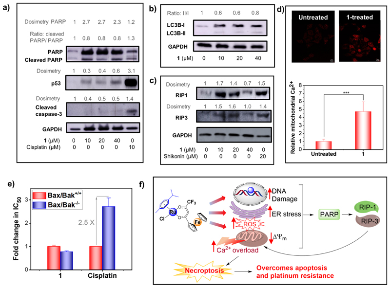
Figure 12.
(a) Western blot analysis of apoptosis-related marker proteins in HeLa cells treated with 1/4 × IC50/24h (10 μM), 1/2 × IC50/24h (20 μM), and IC50/24h (40 μM) concentrations of 1 (24 h compound exposure). (b) Western blot analysis of autophagy marker proteins in HeLa cells treated with concentrations of 1 (24 h compound exposure). (c) Western blot analysis of necroptosis marker proteins in HeLa cells treated with concentrations of 1 (24 h compound exposure). (d) Comparison of the mitochondrial Ca2+ level in untreated or 1-treated (20 μM, 24 h) HeLa cells. (e) Fold change in IC50 of 1 and cisplatin in the wild-type murine embryonic fibroblast (MEF-Bax/Bak+/+) as compared to that in the Bax/ Bak double knocked-out (MEF-Bax/Bak−/−) cell lines. (f) Proposed cellular mechanism of action of 1. * denotes statistical significance (p < 0.05).

