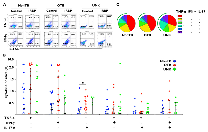
Figure 2.
Interphotoreceptor retinoid-binding protein (IRBP)-specific cytokine responses are comparable between tuberculosis (TB)-immunoreactive and TB-nonreactive phenotypes. Vitreous infiltrated cells from patients with uveitis were stimulated with 10 μg/mL of IRBP1 peptide (IRBP [1-20]) along with anti-CD28 antibody (2 μg/mL) for approximately 14 hours. In the last 8 hours,10 μg/mL Brefeldin A and/or 2 μmol/mL monensin were added. Cells were fixed, stained, and analyzed for tumor necrosis factor-alfa (TNFα), interleukin 17 (IL-17), and interferon-gamma (IFNγ) by flow cytometry. (A) Representative dot plot from 1 patient sample for each of the 3 groups: non-TB control subjects, patients with ocular TB (OTB), and patients with uveitis of unknown origin (UNK). (B) Bar figure representing TNFα, IFNγ, and IL-17 mono- and dual-cytokine responses in each group. (C) Pie chart representing the proportion of TNFα, IFNγ, and IL-17 mono- and dual-cytokine responses in each group. The arcs represent the total polyfunctional component of the antigen-specific response in each group. IRBP1-specific cytokine percentages were subtracted from paired unstimulated samples and the resulting positive cytokines percentages from different groups were compared using the Wilcoxon rank sum test. Data are shown as median ± IQR. P < 0.5 was considered statistically significant. *P ≤ .05; **P ≤ .01; ***P ≤ .001. (Non-TB group, n = 24; OTB group, n = 23; and UNK group, n = 24.)

