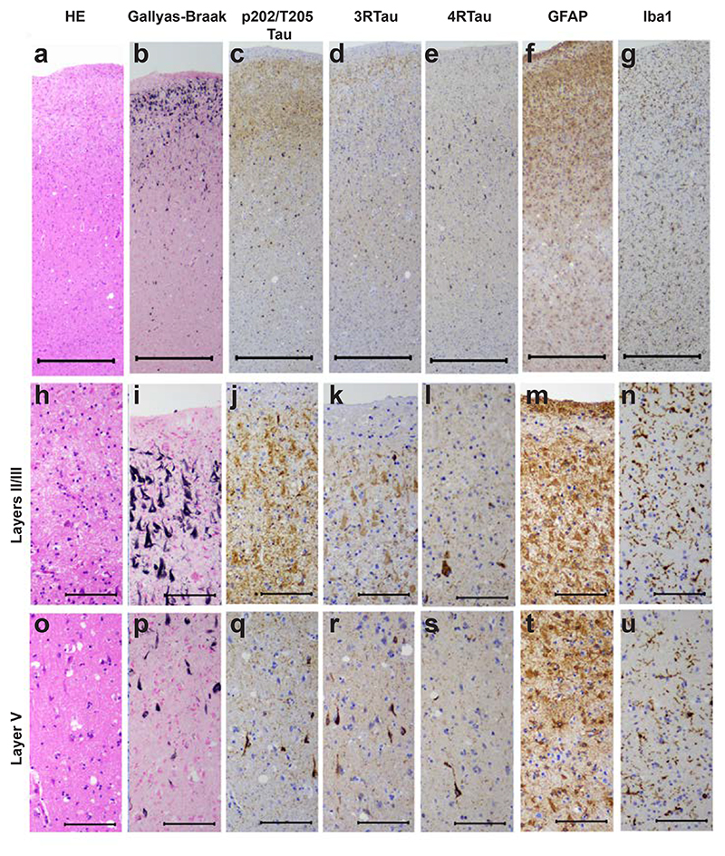
Figure 3. Staining of the superior frontal cortex in vacuolar tauopathy.
Nerve cell loss and gliosis are in seen in layers II/III (a,f,g,h,m,n), where abundant tau-immunoreactive neurofibrillary lesions are in evidence (b,c,d,e,i,j,k,l). Fewer neurofibrillary lesions are seen in layer V (p,q,r,s). Mild vacuolar changes are present in layer V (o). Astrogliosis and microglial changes are most severe in the superficial cortical layers (f,g,m,n). HE staining (a,h,o); Gallyas-Braak silver (b,i,p); pTau (AT8) (c,j,q); 3R Tau (RD3) (d,k,r); 4R Tau (anti-4R) (e,l,s); GFAP (f,m,t); Iba1 (g,n,u). Scale bars: 500 μm (a-g), 100 μm (h-u).

