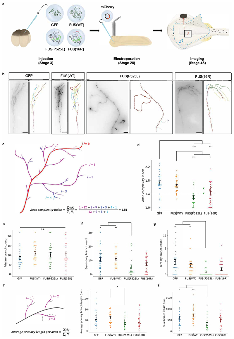
Figure 2. Mutant FUS compromises axonal branching.
a) Schematic of experimental procedure. mRNA encoding only GFP (green) or FUS (grey) fused to GFP is injected at the four-cell stage. At stage 28, axons are sparsely labelled by electroporation, which are then imaged at stage 45. b) Sample images of mCherry-labelled axons expressing different GFP or FUS-GFP constructs. c) Schematic of calculation of axon complexity index (ACI). d) Mutant FUS reduces the axon complexity index. Dashed line indicates value below which axons are considered ‘simple’. e-g) FUS(P52L) reduces the number of higher-order branches. Plots show number of primary, secondary, and tertiary branches per axon respectively. h) FUS(P525L) may reduce average primary branch length. i) FUS(P525L) reduces total branch length. d-i): Number of axons analysed: nGFP=26; nFUS(WT)=25; nFUS(P525L)=21; nFUS(16R)=28. N≥6 replicates for each condition. All conditions were compared pairwise, those that are significantly different are indicated. Kruskal-Wallis tests with Bonferroni correction, error bars indicate standard errors in means.

