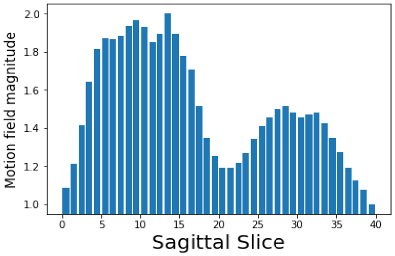
Fig. 5. Normalised motion field magnitude for each sagittal slice in the high-resolution dynamic synthetic MR volumes.
The two peaks correspond to the slices through the left and right lungs, and the right lung shows more significant motion due to the heart obscuring the left lung (as in the images in Fig. 9 the volunteer’s right lung corresponds to the left of the image). Using these values as the dataset weights cn will place more emphasis on the sagittal slices in the centre of the lungs, rather than those around the edge of the torso or around the spine. Here the weights are normalised to have a maximal value of C = 2 and a minimum of 1.

