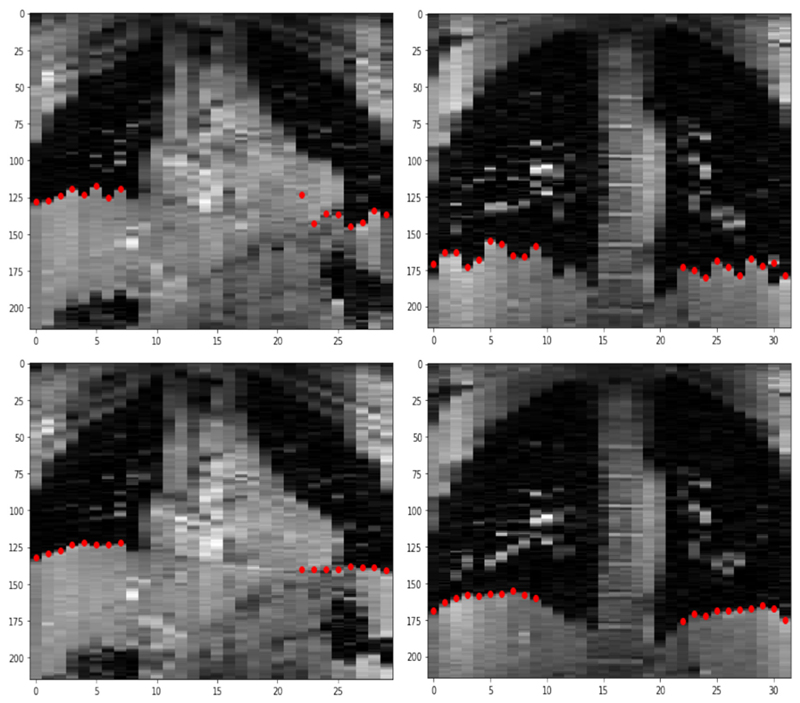
Fig. 8.
Top: Examples from volunteers A (left) and D (right) of a coronal slice through the original unaligned volumes. The sagittal slices are not in consistent motion states resulting in discontinuities in diaphragm positions. Bottom: Examples from a volume reconstructed by stacking sagittal slices aligned by motion state. Estimated diaphragm positions for left and right lungs are shown in red.

