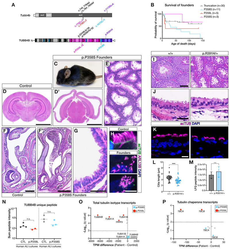
Figure 4. TUBB4B variants caused dominant disease in vivo.
(A) Schematic of patient p.P259L/S (PCD-only, red or magenta), p.P358S (syndromic PCD+LCA, blue) and p.R391H (SND-only, purple) mutations and truncating mutations (p.D249Lfs*29 and p.N256Yfs*32, null alleles (black)) on the mouse Tubb4b mRNA (top) and protein (bottom).
(B-H) (B) Kaplan-Meier graph of founder mice carrying PCD-patient variants which died either spontaneously or were euthanized for health concerns. (C-H) p.P358S founders exhibited hydrocephaly (C,D’), defects in spermatogenesis (E), mucopurulent nasal plugs (F’), and loss of tracheal cilia (G,H). BL/6 age-matched controls (D,F).
(I-M) Tubb4bR391H/+ animals showed no decrease in fitness or survival allowing a line to be established (see also Fig. S9). Males were infertile with defects in spermatogenesis (I). Tubb4bR391H/+ tracheal cilia histology (J), immunofluorescence, length quantified in (L). Mass spectrometry of trachea quantifying TUBB4B levels (M).
(N-P) Nasal epithelial cultures from healthy controls, a PCD-only patient (red, p. P259L) or syndromic PCD+SND patient (blue, p.P358S) were used for mass spectrometry of unique TUBB4B peptides (N) or targeted RNASeq analysis of tubulin (O) or tubulin chaperone (P) transcripts.
Scale bars: 2.5 mm (D), 500 μm (E), 250 μm (F), 100 μm (I), 50 μm (G), 25 μm (J) and 10 μm (H,K). (L, M) Graphic bars: mean ± SEM from N=2 biological replicates, n>20 cells/replicate (L) and N= 3 biological (M) or experimental (N) replicates. Student’s t-test: ns, not significant; ****, p ≤ 0.0001.

