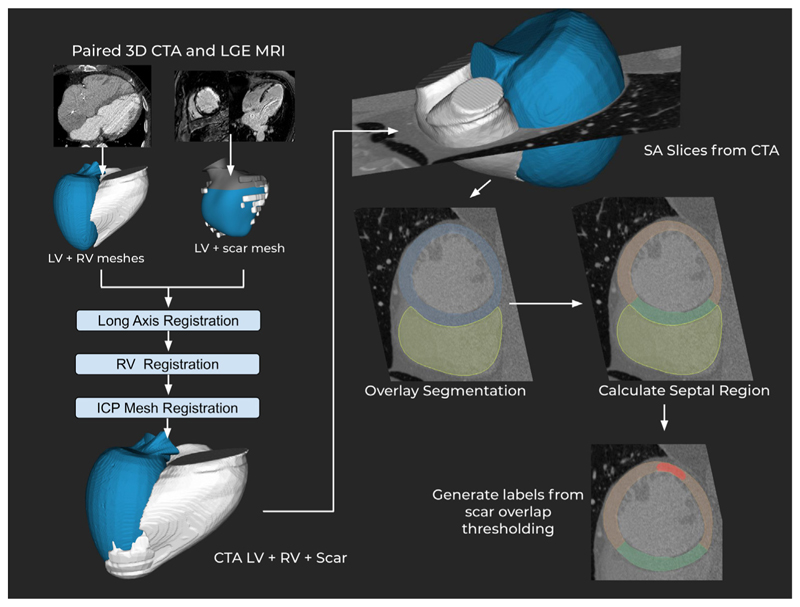
Fig. 1. Data generation process for the septal/lateral scar CTA dataset.
MRI meshes are registered to the CTA meshes in three steps. The meshes are aligned with the CTA DICOM volume, from which SA slices are taken. The septal area is calculated and scar class is determined by a threshold of volume against the volume of the region. ICP: iterative closest point registration.

