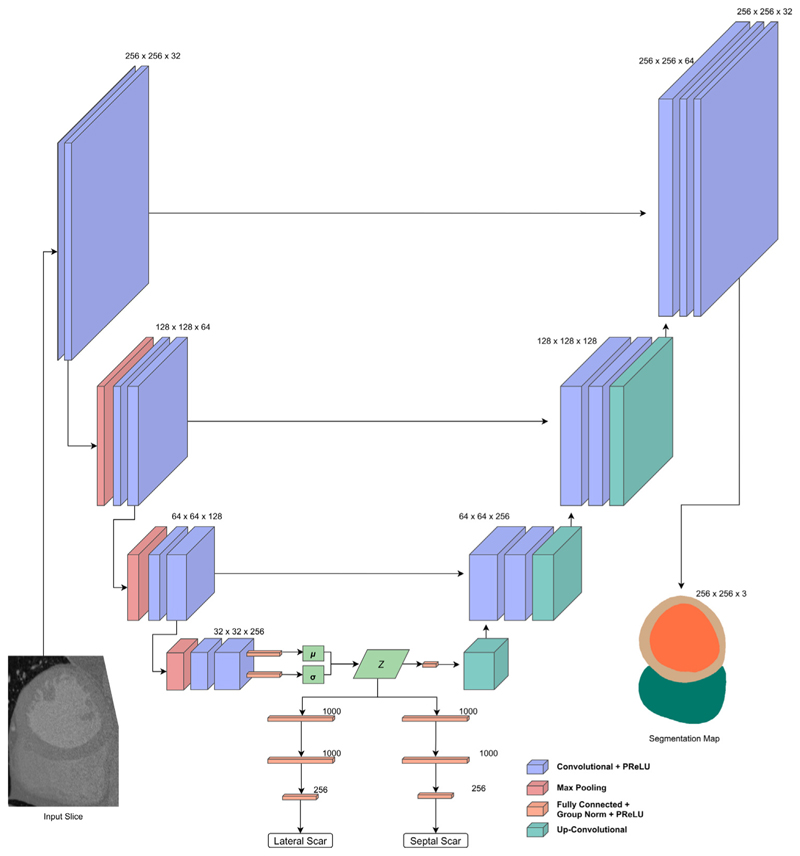
Fig. 3. U-Scar network design.
U-Net style network with convolutional layer blocks as an encoder followed by a de-convolution block decoder with skip connections. The output is a segmentation map of the endocardium, epicardium and RV. The intermediate part of the network is that of a VAE where a latent space is constructed. This latent space is the input to the decoder and a pair of fully connected classification networks predicting scar in either lateral or septal regions.

