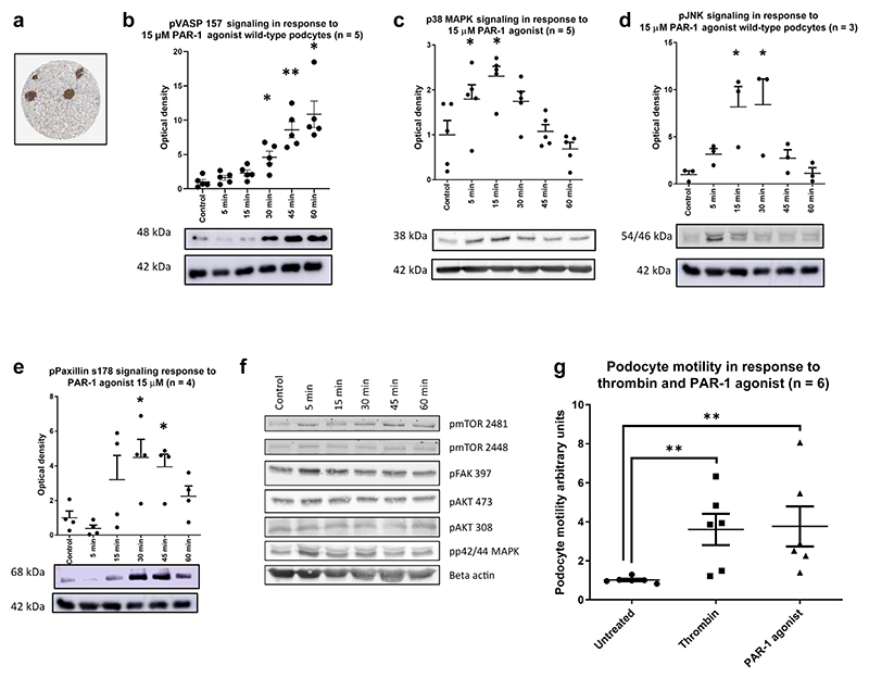
Figure 1. Protease-activatedreceptor 1 (PAR-1) is highly expressed in the podocyte, and its activation is detrimental.
(a) Data from Sigma’s Protein Atlas show an enrichment of PAR-1 expression within the glomeruli of the kidney. All densitometry graphs show the optical density of the band, normalized to the control and corrected by β-actin load control. An example blot is shown beneath each graph. Wild-type conditionally immortalized human podocytes were treated with a PAR-1 agonist at a dose of 15 μM for the indicated time points. There was significant phosphorylation of VASP (b), p38 mitogen-activated protein kinase (MAPK) (c), JNK (d), and Paxillin (e) Bonferroni’s multiple comparison test, *P ≤ 0.05, **P ≤ 0.01, ***P ≤ 0.001, ****P ≤ 0.0001. A range of pathways were interrogated that were not significantly stimulated (f). A wound-healing assay was performed to assess the ability of the PAR-1 agonist to induce a motile phenotype (g). Data shown n = 3 in duplicate, normalized to untreated control. Both the PAR-1 agonist and thrombin treatments significantly increase podocyte motility (1-tailed Mann-Whitney test P = 0.0011 and 0.0022, respectively). pJNK, phospho–c-Jun N-terminal kinase; pVASP, phospho–vasodilator-stimulated phosphoprotein.

