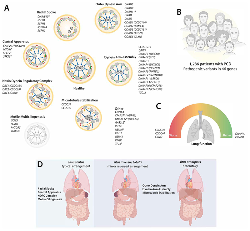
Figure 1. Revealing genotype-phenotype relationships in PCD.
(A) Schematic of motile cilia axoneme cross-section representing ultrastructure required for motility (healthy donor) compared to groups based on structure affected in patients with PCD. 52 PCD genes are listed in italics, with superscript (N) representing near normal ultrastructure. Outer dynein arm (red); inner dynein arm (orange); nexin dynein regulatory complex (green); radial spokes (blue) and central pair apparatus (dark grey). (B) Study overview with number of patients with PCD included from the ERN-LUNG International PCD registry, who carry pathogenic variants in 46 genes. (C) Analysis of large cohorts reveal genotype-phenotype relationships at a gene-level, such as for lung function (FEV1) both as individual data points and longitudinally. Patients with variants in CCDC39, CCDC40 and CCNO have significantly worse lung function (red dial, left) and decline faster than patients with variants in DNAH11 or ODAD1 (green dial, right). (D) In contrast, situs defects are not observed in patients with PCD who have pathogenic variants affecting genes required for motile multiciliogenesis, radial spokes, central apparatus or nexin dynein regulatory complex (blue box, left), in contrast to those affecting formation or function of dynein arms which do show situs inversus totalis or heterotaxy (purple box, right). Elements of figure panels created with BioRender.

